


 Foundational characteristics of cancer include proliferation, angiogenesis, migration, evasion of apoptosis, and cellular immortality. Find key markers for these cellular processes and antibodies to detect them.
 The SUMOplot™ Analysis Program predicts and scores sumoylation sites in your protein. SUMOylation is a post-translational modification involved in various cellular processes, such as nuclear-cytosolic transport, transcriptional regulation, apoptosis, protein stability, response to stress, and progression through the cell cycle.
 The Autophagy Receptor Motif Plotter predicts and scores autophagy receptor binding sites in your protein. Identifying proteins connected to this pathway is critical to understanding the role of autophagy in physiological as well as pathological processes such as development, differentiation, neurodegenerative diseases, stress, infection, and cancer.
Anti-APOBEC3G Antibody Picoband™ (monoclonal, 6C2)

- SPECIFICATION
- CITATIONS
- PROTOCOLS
- BACKGROUND

Application
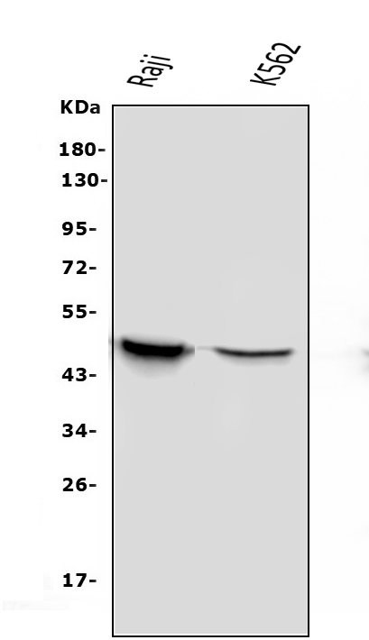
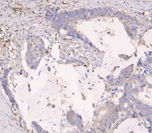
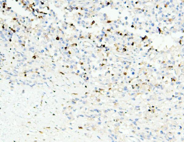
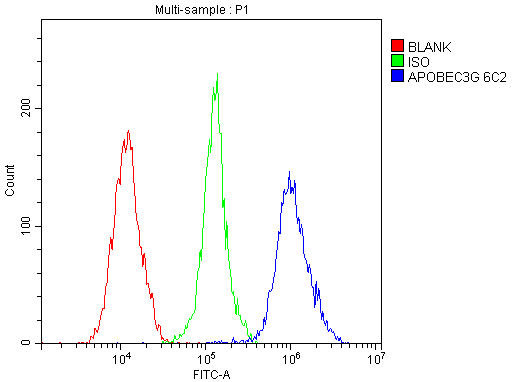
| WB, IHC, IF, ICC, FC |
|---|---|
| Primary Accession | Q9HC16 |
| Host | Mouse |
| Isotype | Mouse IgG1 |
| Reactivity | Human |
| Clonality | Monoclonal |
| Format | Lyophilized |
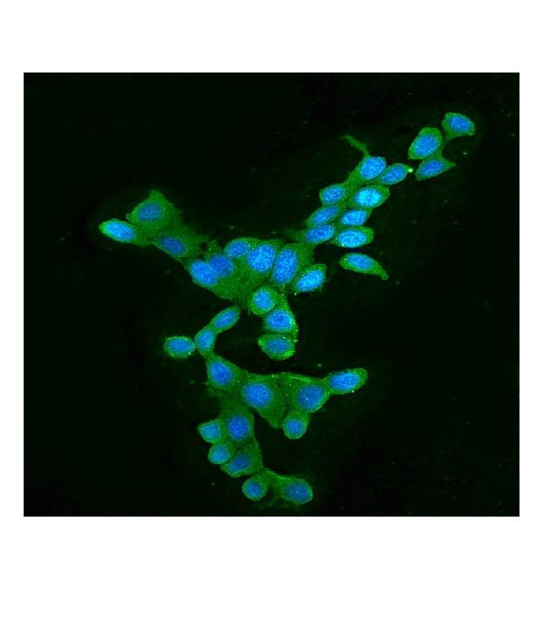
| Description | Anti-APOBEC3G Antibody Picoband™ (monoclonal, 6C2) . Tested in Flow Cytometry, IF, IHC, ICC, WB applications. This antibody reacts with Human. |
| Reconstitution | Add 0.2ml of distilled water will yield a concentration of 500 µg/ml. |
| Gene ID | 60489 |
|---|---|
| Other Names | DNA dC->dU-editing enzyme APOBEC-3G, 3.5.4.38, APOBEC-related cytidine deaminase, APOBEC-related protein, ARCD, APOBEC-related protein 9, ARP-9, CEM-15, CEM15, Deoxycytidine deaminase, A3G, APOBEC3G |
| Calculated MW | 46 kDa |
| Application Details | Western blot, 0.1-0.5 µg/ml Immunohistochemistry (Paraffin-embedded Section), 0.5-1 µg/ml Immunocytochemistry/Immunofluorescence, 2 µg/ml Flow Cytometry, 1-3 µg/1x10^6 cells |
| Subcellular Localization | Nucleus. Cytoplasm. P-body. |
| Tissue Specificity | Expressed in spleen, testes, ovary and peripheral blood leukocytes and CD4+ lymphocytes. Also expressed in non-permissive peripheral blood mononuclear cells, and several tumor cell lines; no expression detected in permissive lymphoid and non-lymphoid cell lines. Exists only in the LMM form in peripheral blood-derived resting CD4 T-cells and monocytes, both of which are refractory to HIV-1 infection. LMM is converted to a HMM complex when resting CD4 T-cells are activated or when monocytes are induced to differentiate into macrophages. This change correlates with increased susceptibility of these cells to HIV-1 infection. |
| Contents | Each vial contains 4mg Trehalose, 0.9mg NaCl, 0.2mg Na2HPO4, 0.05mg NaN3. |
| Clone Names | Clone: 6C2 |
| Immunogen | E.coli-derived human APOBEC3G recombinant protein (Position: E191-N384). |
| Cross Reactivity | No cross-reactivity with other proteins. |
| Storage | Store at -20˚C for one year from date of receipt. After reconstitution, at 4˚C for one month. It can also be aliquotted and stored frozen at -20˚C for six months. Avoid repeated freeze-thaw cycles. |
| Name | APOBEC3G {ECO:0000303|PubMed:14557625, ECO:0000312|HGNC:HGNC:17357} |
|---|---|
| Function | DNA deaminase (cytidine deaminase) which acts as an inhibitor of retrovirus replication and retrotransposon mobility via deaminase- dependent and -independent mechanisms (PubMed:12808465, PubMed:16527742, PubMed:17121840, PubMed:18288108, PubMed:18849968, PubMed:19153609, PubMed:21123384, PubMed:22791714, PubMed:25542899). Exhibits potent antiviral activity against Vif-deficient HIV-1 (PubMed:12167863, PubMed:12859895, PubMed:14557625, PubMed:20219927, PubMed:21835787, PubMed:22807680, PubMed:22915799, PubMed:23097438, PubMed:23152537, PubMed:31397674). After the penetration of retroviral nucleocapsids into target cells of infection and the initiation of reverse transcription, it can induce the conversion of cytosine to uracil in the minus-sense single-strand viral DNA, leading to G-to-A hypermutations in the subsequent plus-strand viral DNA (PubMed:12808465, PubMed:12808466, PubMed:12809610, PubMed:12970355, PubMed:14528300, PubMed:22807680). The resultant detrimental levels of mutations in the proviral genome, along with a deamination-independent mechanism that works prior to the proviral integration, together exert efficient antiretroviral effects in infected target cells (PubMed:12808465, PubMed:12808466, PubMed:12809610, PubMed:12970355, PubMed:14528300). Selectively targets single-stranded DNA and does not deaminate double-stranded DNA or single- or double-stranded RNA (PubMed:12808465, PubMed:12809610, PubMed:12970355, PubMed:14528300). Exhibits antiviral activity also against simian immunodeficiency viruses (SIVs), hepatitis B virus (HBV), equine infectious anemia virus (EIAV), xenotropic MuLV-related virus (XMRV) and simian foamy virus (SFV) (PubMed:15031497, PubMed:16378963, PubMed:18448976, PubMed:19458006, PubMed:20335265). May inhibit the mobility of LTR and non-LTR retrotransposons (PubMed:16527742). |
| Cellular Location | Cytoplasm. Nucleus Cytoplasm, P-body. Note=Mainly cytoplasmic (PubMed:16527742, PubMed:16699599, PubMed:21835787). Small amount are found in the nucleus (PubMed:18667511). During HIV-1 infection, virion-encapsidated in absence of HIV-1 Vif (PubMed:12859895) |
| Tissue Location | Expressed in spleen, testes, ovary and peripheral blood leukocytes and CD4+ lymphocytes. Also expressed in non-permissive peripheral blood mononuclear cells, and several tumor cell lines; no expression detected in permissive lymphoid and non-lymphoid cell lines Exists only in the LMM form in peripheral blood-derived resting CD4 T- cells and monocytes, both of which are refractory to HIV-1 infection LMM is converted to a HMM complex when resting CD4 T-cells are activated or when monocytes are induced to differentiate into macrophages. This change correlates with increased susceptibility of these cells to HIV-1 infection. |

Thousands of laboratories across the world have published research that depended on the performance of antibodies from Abcepta to advance their research. Check out links to articles that cite our products in major peer-reviewed journals, organized by research category.
info@abcepta.com, and receive a free "I Love Antibodies" mug.

Provided below are standard protocols that you may find useful for product applications.
Background
APOBEC3G (apolipoprotein B mRNA editing enzyme, catalytic polypeptide-like 3G) is a human enzyme encoded by the APOBEC3G gene. This gene is a member of the cytidine deaminase gene family. It is one of seven related genes or pseudogenes found in a cluster, thought to result from gene duplication, on chromosome 22. Members of the cluster encode proteins that are structurally and functionally related to the C to U RNA-editing cytidine deaminase APOBEC1. It is thought that the proteins may be RNA editing enzymes and have roles in growth or cell cycle control. The protein encoded by this gene has been found to be a specific inhibitor of human immunodeficiency virus-1 (HIV-1) infectivity.
If you have used an Abcepta product and would like to share how it has performed, please click on the "Submit Review" button and provide the requested information. Our staff will examine and post your review and contact you if needed.
If you have any additional inquiries please email technical services at tech@abcepta.com.
Ordering Information
Other Products
Shipping Information
