


 Foundational characteristics of cancer include proliferation, angiogenesis, migration, evasion of apoptosis, and cellular immortality. Find key markers for these cellular processes and antibodies to detect them.
 The SUMOplot™ Analysis Program predicts and scores sumoylation sites in your protein. SUMOylation is a post-translational modification involved in various cellular processes, such as nuclear-cytosolic transport, transcriptional regulation, apoptosis, protein stability, response to stress, and progression through the cell cycle.
 The Autophagy Receptor Motif Plotter predicts and scores autophagy receptor binding sites in your protein. Identifying proteins connected to this pathway is critical to understanding the role of autophagy in physiological as well as pathological processes such as development, differentiation, neurodegenerative diseases, stress, infection, and cancer.
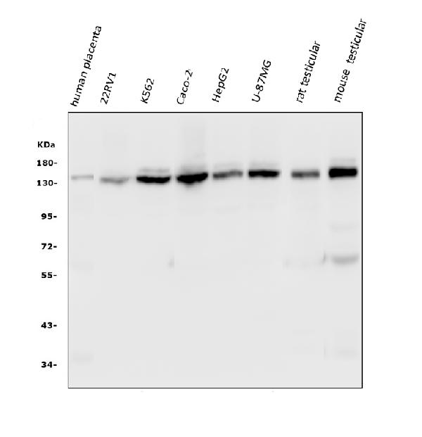
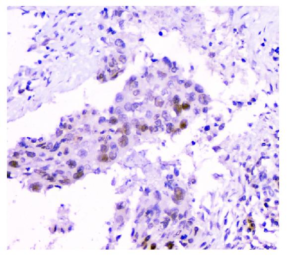
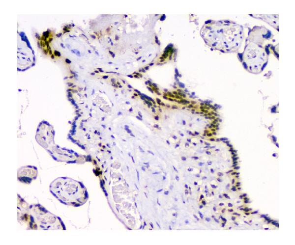
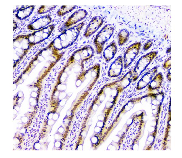
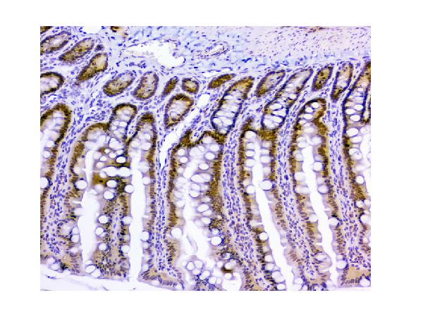
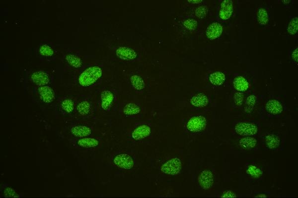
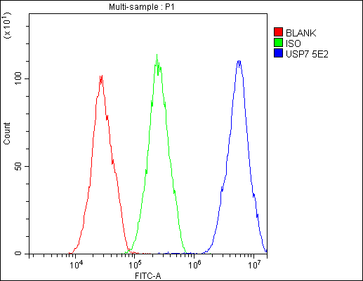
Anti-HAUSP/USP7 Antibody Picoband™ (monoclonal, 5E2)
- SPECIFICATION
- CITATIONS
- PROTOCOLS
- BACKGROUND

Application
| WB, IHC, IF, ICC, FC |
|---|---|
| Primary Accession | Q93009 |
| Host | Mouse |
| Isotype | Mouse IgG2b |
| Reactivity | Rat, Human, Mouse |
| Clonality | Monoclonal |
| Format | Lyophilized |
| Description | Anti-HAUSP/USP7 Antibody Picoband™ (monoclonal, 5E2) . Tested in Flow Cytometry, IF, IHC, ICC, WB applications. This antibody reacts with Human, Mouse, Rat. |
| Reconstitution | Add 0.2ml of distilled water will yield a concentration of 500 µg/ml. |
| Gene ID | 7874 |
|---|---|
| Other Names | Ubiquitin carboxyl-terminal hydrolase 7, 3.4.19.12, Deubiquitinating enzyme 7, Herpesvirus-associated ubiquitin-specific protease, Ubiquitin thioesterase 7, Ubiquitin-specific-processing protease 7, USP7 {ECO:0000303|PubMed:12093161, ECO:0000312|HGNC:HGNC:12630} |
| Calculated MW | 140 kDa |
| Application Details | Western blot, 0.1-0.5 µg/ml Immunohistochemistry (Paraffin-embedded Section), 0.5-1 µg/ml Immunocytochemistry/Immunofluorescence, 2 µg/ml Flow Cytometry, 1-3 µg/1x10^6 cells |
| Subcellular Localization | Nucleus. PML body. Cytoplasm. Chromosome. |
| Tissue Specificity | Widely expressed. Overexpressed in prostate cancer. |
| Contents | Each vial contains 4mg Trehalose, 0.9mg NaCl, 0.2mg Na2HPO4, 0.05mg NaN3. |
| Clone Names | Clone: Clone: 5E2 |
| Immunogen | E.coli-derived human HAUSP/USP7 recombinant protein (Position: L258-D483). |
| Cross Reactivity | No cross-reactivity with other proteins. |
| Storage | Store at -20˚C for one year from date of receipt. After reconstitution, at 4˚C for one month. It can also be aliquotted and stored frozen at -20˚C for six months. Avoid repeated freeze-thaw cycles. |
| Name | USP7 {ECO:0000303|PubMed:12093161, ECO:0000312|HGNC:HGNC:12630} |
|---|---|
| Function | Hydrolase that deubiquitinates target proteins such as FOXO4, DEPTOR, KAT5, p53/TP53, MDM2, ERCC6, DNMT1, UHRF1, PTEN, KMT2E/MLL5 and DAXX (PubMed:11923872, PubMed:15053880, PubMed:16964248, PubMed:18716620, PubMed:25283148, PubMed:25865756, PubMed:26678539, PubMed:28655758, PubMed:35216969). Together with DAXX, prevents MDM2 self-ubiquitination and enhances the E3 ligase activity of MDM2 towards p53/TP53, thereby promoting p53/TP53 ubiquitination and proteasomal degradation (PubMed:15053880, PubMed:16845383, PubMed:18566590, PubMed:20153724). Deubiquitinates p53/TP53, preventing degradation of p53/TP53, and enhances p53/TP53-dependent transcription regulation, cell growth repression and apoptosis (PubMed:25283148). Deubiquitinates p53/TP53 and MDM2 and strongly stabilizes p53/TP53 even in the presence of excess MDM2, and also induces p53/TP53-dependent cell growth repression and apoptosis (PubMed:11923872, PubMed:26786098). Deubiquitination of FOXO4 in presence of hydrogen peroxide is not dependent on p53/TP53 and inhibits FOXO4-induced transcriptional activity (PubMed:16964248). In association with DAXX, is involved in the deubiquitination and translocation of PTEN from the nucleus to the cytoplasm, both processes that are counteracted by PML (PubMed:18716620). Deubiquitinates KMT2E/MLL5 preventing KMT2E/MLL5 proteasomal-mediated degradation (PubMed:26678539). Involved in cell proliferation during early embryonic development. Involved in transcription-coupled nucleotide excision repair (TC-NER) in response to UV damage: recruited to DNA damage sites following interaction with KIAA1530/UVSSA and promotes deubiquitination of ERCC6, preventing UV- induced degradation of ERCC6 (PubMed:22466611, PubMed:22466612). Involved in maintenance of DNA methylation via its interaction with UHRF1 and DNMT1: acts by mediating deubiquitination of UHRF1 and DNMT1, preventing their degradation and promoting DNA methylation by DNMT1 (PubMed:21745816, PubMed:22411829). Deubiquitinates alkylation repair enzyme ALKBH3. OTUD4 recruits USP7 and USP9X to stabilize ALKBH3, thereby promoting the repair of alkylated DNA lesions (PubMed:25944111). Acts as a chromatin regulator via its association with the Polycomb group (PcG) multiprotein PRC1-like complex; may act by deubiquitinating components of the PRC1-like complex (PubMed:20601937). Able to mediate deubiquitination of histone H2B; it is however unsure whether this activity takes place in vivo (PubMed:20601937). Exhibits a preference towards 'Lys-48'-linked ubiquitin chains (PubMed:22689415). Increases regulatory T-cells (Treg) suppressive capacity by deubiquitinating and stabilizing the transcription factor FOXP3 which is crucial for Treg cell function (PubMed:23973222). Plays a role in the maintenance of the circadian clock periodicity via deubiquitination and stabilization of the CRY1 and CRY2 proteins (PubMed:27123980). Deubiquitinates REST, thereby stabilizing REST and promoting the maintenance of neural progenitor cells (PubMed:21258371). Deubiquitinates SIRT7, inhibiting SIRT7 histone deacetylase activity and regulating gluconeogenesis (PubMed:28655758). Involved in the regulation of WASH-dependent actin polymerization at the surface of endosomes and the regulation of endosomal protein recycling (PubMed:26365382). It maintains optimal WASH complex activity and precise F-actin levels via deubiquitination of TRIM27 and WASHC1 (PubMed:26365382). Mediates the deubiquitination of phosphorylated DEPTOR, promoting its stability and leading to decreased mTORC1 signaling (PubMed:35216969). |
| Cellular Location | Nucleus. Cytoplasm Nucleus, PML body. Chromosome. Note=Present in a minority of ND10 nuclear bodies. Association with ICP0/VMW110 at early times of infection leads to an increased proportion of USP7-containing ND10 Colocalizes with ATXN1 in the nucleus. Colocalized with DAXX in speckled structures. Colocalized with PML and PTEN in promyelocytic leukemia protein (PML) nuclear bodies |
| Tissue Location | Expressed in neural progenitor cells (at protein level) (PubMed:21258371). Widely expressed. Overexpressed in prostate cancer. |

Thousands of laboratories across the world have published research that depended on the performance of antibodies from Abcepta to advance their research. Check out links to articles that cite our products in major peer-reviewed journals, organized by research category.
info@abcepta.com, and receive a free "I Love Antibodies" mug.

Provided below are standard protocols that you may find useful for product applications.
Background
Ubiquitin-specific-processing protease 7 (USP7), also known as ubiquitin carboxyl-terminal hydrolase 7 or herpesvirus-associated ubiquitin-specific protease (HAUSP), is an enzyme that in humans is encoded by the USP7 gene. The protein encoded by this gene belongs to the peptidase C19 family, which includes ubiquitinyl hydrolases. This protein deubiquitinates target proteins such as p53 (a tumor suppressor protein) and WASH (essential for endosomal protein recycling), and regulates their activities by counteracting the opposing ubiquitin ligase activity of proteins such as HDM2 and TRIM27, involved in the respective process. Mutations in this gene have been implicated in a neurodevelopmental disorder.
If you have used an Abcepta product and would like to share how it has performed, please click on the "Submit Review" button and provide the requested information. Our staff will examine and post your review and contact you if needed.
If you have any additional inquiries please email technical services at tech@abcepta.com.
Ordering Information
Other Products
Shipping Information
