


 Foundational characteristics of cancer include proliferation, angiogenesis, migration, evasion of apoptosis, and cellular immortality. Find key markers for these cellular processes and antibodies to detect them.
 The SUMOplot™ Analysis Program predicts and scores sumoylation sites in your protein. SUMOylation is a post-translational modification involved in various cellular processes, such as nuclear-cytosolic transport, transcriptional regulation, apoptosis, protein stability, response to stress, and progression through the cell cycle.
 The Autophagy Receptor Motif Plotter predicts and scores autophagy receptor binding sites in your protein. Identifying proteins connected to this pathway is critical to understanding the role of autophagy in physiological as well as pathological processes such as development, differentiation, neurodegenerative diseases, stress, infection, and cancer.
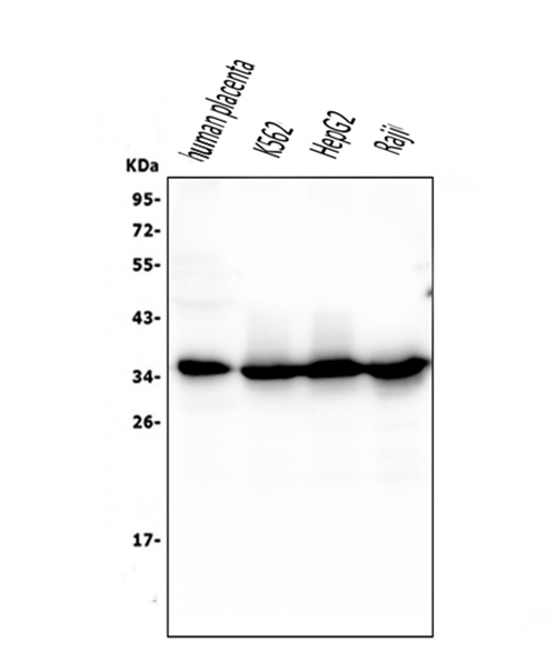
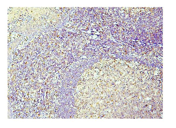
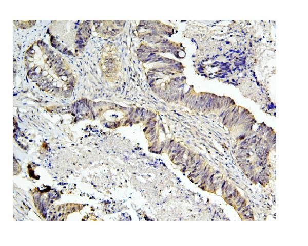
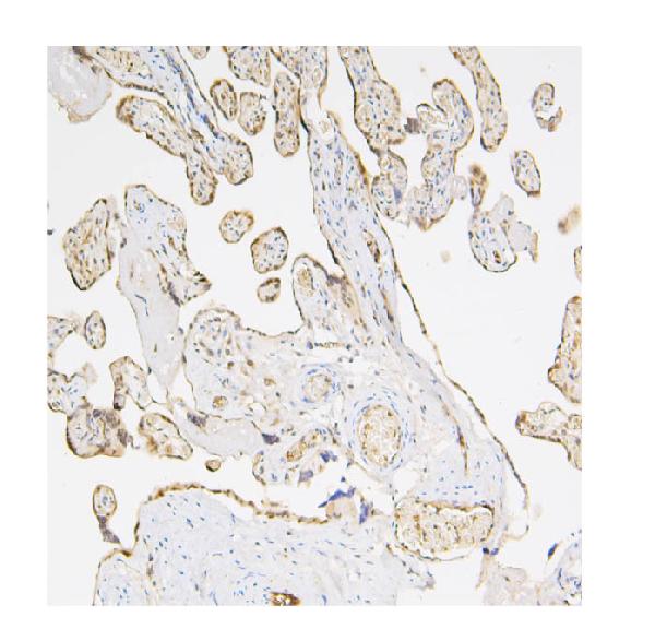
Anti-Caspase-3 Antibody Picoband™ (monoclonal, 8B6)
- SPECIFICATION
- CITATIONS
- PROTOCOLS
- BACKGROUND

Application
| WB, IHC, FC |
|---|---|
| Primary Accession | P42574 |
| Host | Mouse |
| Isotype | Mouse IgG1 |
| Reactivity | Human |
| Clonality | Monoclonal |
| Format | Lyophilized |
| Description | Anti-Caspase-3 Antibody Picoband™ (monoclonal, 8B6) . Tested in Flow Cytometry, IHC, WB applications. This antibody reacts with Human. |
| Reconstitution | Add 0.2ml of distilled water will yield a concentration of 500 µg/ml. |
| Gene ID | 836 |
|---|---|
| Other Names | Caspase-3, CASP-3, 3.4.22.56, Apopain, Cysteine protease CPP32, CPP-32, Protein Yama, SREBP cleavage activity 1, SCA-1, Caspase-3 subunit p17, Caspase-3 subunit p12, CASP3, CPP32 {ECO:0000303|PubMed:7983002} |
| Calculated MW | 35 kDa |
| Application Details | Western blot, 0.1-0.5 µg/ml Immunohistochemistry (Paraffin-embedded Section), 0.5-1 µg/ml Flow Cytometry, 1-3 µg/1x10^6 cells |
| Subcellular Localization | Cytoplasm. |
| Tissue Specificity | Highly expressed in lung, spleen, heart, liver and kidney. Moderate levels in brain and skeletal muscle, and low in testis. Also found in many cell lines, highest expression in cells of the immune system. |
| Contents | Each vial contains 4mg Trehalose, 0.9mg NaCl, 0.2mg Na2HPO4, 0.05mg NaN3. |
| Clone Names | Clone: 8B6 |
| Immunogen | E.coli-derived human Caspase-3 recombinant protein (Position: T67-D175). Human Caspase-3 shares 86% and 90% amino acid (aa) sequence identity with mouse and rat Caspase-3, respectively. |
| Cross Reactivity | No cross-reactivity with other proteins. |
| Storage | Store at -20˚C for one year from date of receipt. After reconstitution, at 4˚C for one month. It can also be aliquotted and stored frozen at -20˚C for six months. Avoid repeated freeze-thaw cycles. |
| Name | CASP3 |
|---|---|
| Synonyms | CPP32 {ECO:0000303|PubMed:7983002} |
| Function | Thiol protease that acts as a major effector caspase involved in the execution phase of apoptosis (PubMed:18723680, PubMed:20566630, PubMed:23650375, PubMed:35338844, PubMed:35446120, PubMed:7596430). Following cleavage and activation by initiator caspases (CASP8, CASP9 and/or CASP10), mediates execution of apoptosis by catalyzing cleavage of many proteins (PubMed:18723680, PubMed:20566630, PubMed:23650375, PubMed:7596430). At the onset of apoptosis, it proteolytically cleaves poly(ADP-ribose) polymerase PARP1 at a '216-Asp-|-Gly-217' bond (PubMed:10497198, PubMed:16374543, PubMed:7596430, PubMed:7774019). Cleaves and activates sterol regulatory element binding proteins (SREBPs) between the basic helix-loop-helix leucine zipper domain and the membrane attachment domain (By similarity). Cleaves and activates caspase-6, -7 and -9 (CASP6, CASP7 and CASP9, respectively) (PubMed:7596430). Cleaves and inactivates interleukin-18 (IL18) (PubMed:37993714, PubMed:9334240). Involved in the cleavage of huntingtin (PubMed:8696339). Triggers cell adhesion in sympathetic neurons through RET cleavage (PubMed:21357690). Cleaves and inhibits serine/threonine-protein kinase AKT1 in response to oxidative stress (PubMed:23152800). Acts as an inhibitor of type I interferon production during virus-induced apoptosis by mediating cleavage of antiviral proteins CGAS, IRF3 and MAVS, thereby preventing cytokine overproduction (PubMed:30878284). Also involved in pyroptosis by mediating cleavage and activation of gasdermin-E (GSDME) (PubMed:35338844, PubMed:35446120). Cleaves XRCC4 and phospholipid scramblase proteins XKR4, XKR8 and XKR9, leading to promote phosphatidylserine exposure on apoptotic cell surface (PubMed:23845944, PubMed:33725486). Cleaves BIRC6 following inhibition of BIRC6-caspase binding by DIABLO/SMAC (PubMed:36758104, PubMed:36758106). |
| Cellular Location | Cytoplasm. |
| Tissue Location | Highly expressed in lung, spleen, heart, liver and kidney. Moderate levels in brain and skeletal muscle, and low in testis. Also found in many cell lines, highest expression in cells of the immune system. |

Thousands of laboratories across the world have published research that depended on the performance of antibodies from Abcepta to advance their research. Check out links to articles that cite our products in major peer-reviewed journals, organized by research category.
info@abcepta.com, and receive a free "I Love Antibodies" mug.

Provided below are standard protocols that you may find useful for product applications.
Background
Caspase 3 is a caspase protein which interacts with Survivin, XIAP, CFLAR, Caspase 8, HCLS1, Deleted in Colorectal Cancer, TRAF3 and GroEL. This gene which is located on 4q35 encodes a protein that is a member of the cysteine-aspartic acid protease (caspase) family. Sequential activation of caspases plays a central role in the execution-phase of cell apoptosis. Caspases exist as inactive proenzymes that undergo proteolytic processing at conserved aspartic residues to produce two subunits, large and small, that dimerize to form the active enzyme. It is the predominant caspase involved in the cleavage of amyloid-beta 4A precursor protein, which is associated with neuronal death in Alzheimer's disease. And the caspase-3 activation in heart failure sequentially cleaves SRF and generates a truncated SRF that appears to function as a dominant-negative transcription factor. Additionally, the caspase-3 influence on bone mineral density should be considered in any in vivo application of caspase-3 inhibitors to the treatment of human disease. In erythroid precursors undergoing terminal differentiation, Hsp70 prevents active CASP3 from cleaving GATA1 and inducing apoptosis.
If you have used an Abcepta product and would like to share how it has performed, please click on the "Submit Review" button and provide the requested information. Our staff will examine and post your review and contact you if needed.
If you have any additional inquiries please email technical services at tech@abcepta.com.
Ordering Information
Other Products
Shipping Information
