


 Foundational characteristics of cancer include proliferation, angiogenesis, migration, evasion of apoptosis, and cellular immortality. Find key markers for these cellular processes and antibodies to detect them.
 The SUMOplot™ Analysis Program predicts and scores sumoylation sites in your protein. SUMOylation is a post-translational modification involved in various cellular processes, such as nuclear-cytosolic transport, transcriptional regulation, apoptosis, protein stability, response to stress, and progression through the cell cycle.
 The Autophagy Receptor Motif Plotter predicts and scores autophagy receptor binding sites in your protein. Identifying proteins connected to this pathway is critical to understanding the role of autophagy in physiological as well as pathological processes such as development, differentiation, neurodegenerative diseases, stress, infection, and cancer.
Anti-GPX1 Antibody Picoband™ (monoclonal, 8B10)
- SPECIFICATION
- CITATIONS
- PROTOCOLS
- BACKGROUND

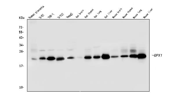
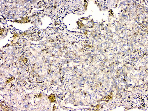
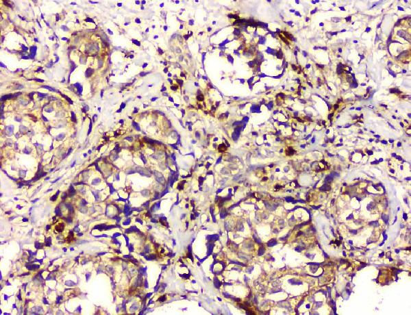
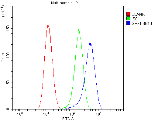
Application
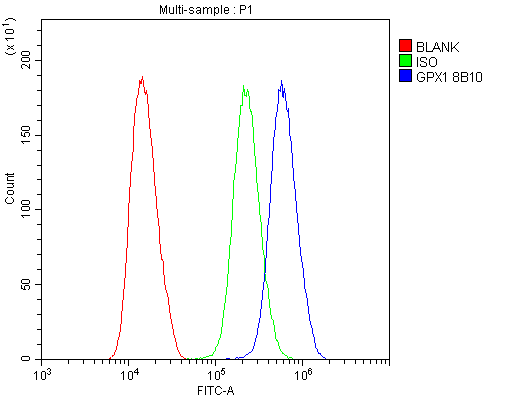
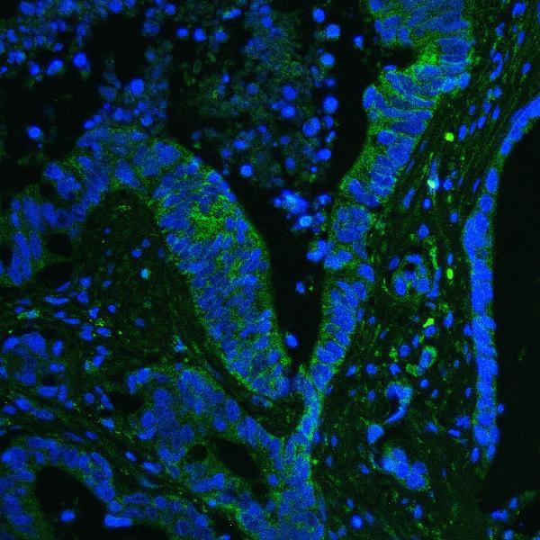
| WB, IHC, IF, ICC, FC |
|---|---|
| Primary Accession | P07203 |
| Host | Mouse |
| Isotype | Mouse IgG2b |
| Reactivity | Rat, Human, Mouse |
| Clonality | Monoclonal |
| Format | Lyophilized |
| Description | Anti-GPX1 Antibody Picoband™ (monoclonal, 8B10) . Tested in Flow Cytometry, IF, IHC, ICC, WB applications. This antibody reacts with Human, Mouse, Rat. |
| Reconstitution | Add 0.2ml of distilled water will yield a concentration of 500 µg/ml. |
| Gene ID | 2876 |
|---|---|
| Other Names | Glutathione peroxidase 1, GPx-1, GSHPx-1, 1.11.1.9, Cellular glutathione peroxidase, Phospholipid-hydroperoxide glutathione peroxidase GPX1, 1.11.1.12, GPX1 (HGNC:4553) |
| Calculated MW | 22 kDa |
| Application Details | Western blot, 0.1-0.5 µg/ml Immunohistochemistry (Paraffin-embedded Section), 0.5-1 µg/ml Immunocytochemistry/Immunofluorescence, 5 µg/ml Flow Cytometry, 1-3 µg/1x10^6 cells |
| Subcellular Localization | Cytoplasm |
| Contents | Each vial contains 4mg Trehalose, 0.9mg NaCl, 0.2mg Na2HPO4, 0.05mg NaN3. |
| Clone Names | Clone: 8B10 |
| Immunogen | A synthetic peptide corresponding to a sequence in the middle region of human GPX1, different from the related mouse sequence by six amino acids and from the related rat sequence by five amino acids. |
| Cross Reactivity | No cross-reactivity with other proteins. |
| Storage | Store at -20˚C for one year from date of receipt. After reconstitution, at 4˚C for one month. It can also be aliquotted and stored frozen at -20˚C for six months. Avoid repeated freeze-thaw cycles. |
| Name | GPX1 (HGNC:4553) |
|---|---|
| Function | Catalyzes the reduction of hydroperoxides in a glutathione- dependent manner thus regulating cellular redox homeostasis (PubMed:11115402, PubMed:36608588). Can reduce small soluble hydroperoxides such as H2O2, cumene hydroperoxide and tert-butyl hydroperoxide, as well as several fatty acid-derived hydroperoxides (PubMed:11115402, PubMed:36608588). In platelets catalyzes the reduction of 12-hydroperoxyeicosatetraenoic acid, the primary product of the arachidonate 12-lipoxygenase pathway (PubMed:11115402). |
| Cellular Location | Cytoplasm {ECO:0000250|UniProtKB:P11352}. Mitochondrion {ECO:0000250|UniProtKB:P11352} |
| Tissue Location | Expressed in platelets (at protein level). |

Thousands of laboratories across the world have published research that depended on the performance of antibodies from Abcepta to advance their research. Check out links to articles that cite our products in major peer-reviewed journals, organized by research category.
info@abcepta.com, and receive a free "I Love Antibodies" mug.

Provided below are standard protocols that you may find useful for product applications.
Background
Glutathione peroxidase 1, also known as, GPX-1 is an enzyme that in humans is encoded by the GPX1 gene. It is mapped to 3p21.31. This gene encodes a member of the glutathione peroxidase family, consisting of eight known glutathione peroxidases (Gpx1-8) in humans. Glutathione peroxidase functions in the detoxification of hydrogen peroxide, and is one of the most important antioxidant enzymes in humans. It has been reported that the protein encoded by this gene protects from CD95-induced apoptosis in cultured breast cancer cells and inhibits 5-lipoxygenase in blood cells, and its overexpression delays endothelial cell growth and increases resistance to toxic challenges. GPX1 is one of only a few proteins known in higher vertebrates to contain selenocysteine, which occurs at the active site of glutathione peroxidase and is coded by the nonsense (stop) codon TGA.
If you have used an Abcepta product and would like to share how it has performed, please click on the "Submit Review" button and provide the requested information. Our staff will examine and post your review and contact you if needed.
If you have any additional inquiries please email technical services at tech@abcepta.com.
Ordering Information
Other Products
Shipping Information
