


 Foundational characteristics of cancer include proliferation, angiogenesis, migration, evasion of apoptosis, and cellular immortality. Find key markers for these cellular processes and antibodies to detect them.
 The SUMOplot™ Analysis Program predicts and scores sumoylation sites in your protein. SUMOylation is a post-translational modification involved in various cellular processes, such as nuclear-cytosolic transport, transcriptional regulation, apoptosis, protein stability, response to stress, and progression through the cell cycle.
 The Autophagy Receptor Motif Plotter predicts and scores autophagy receptor binding sites in your protein. Identifying proteins connected to this pathway is critical to understanding the role of autophagy in physiological as well as pathological processes such as development, differentiation, neurodegenerative diseases, stress, infection, and cancer.
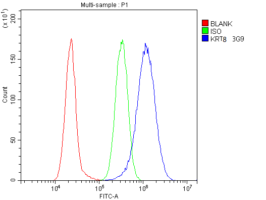
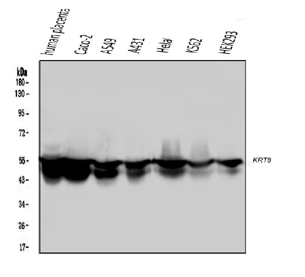
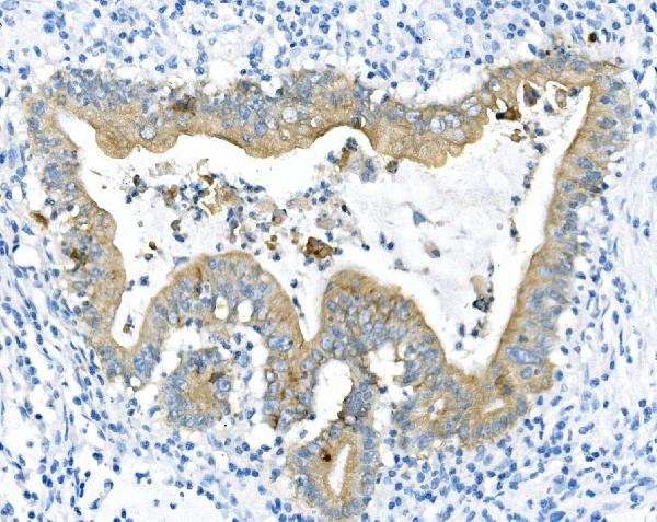
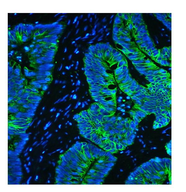
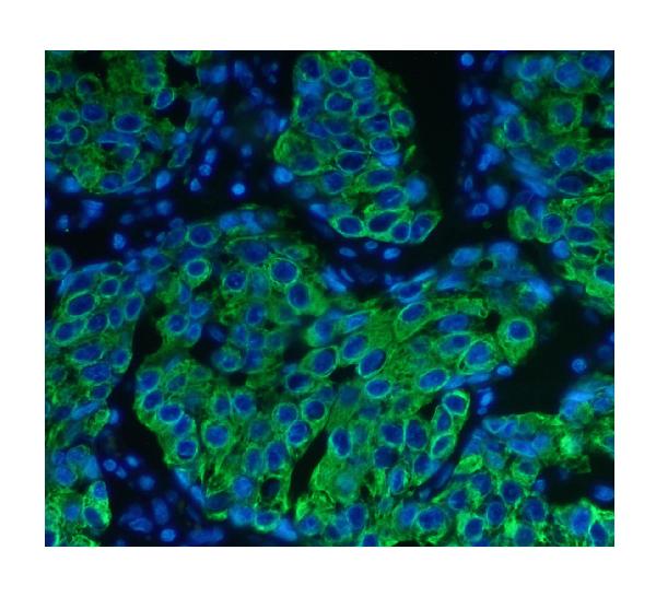
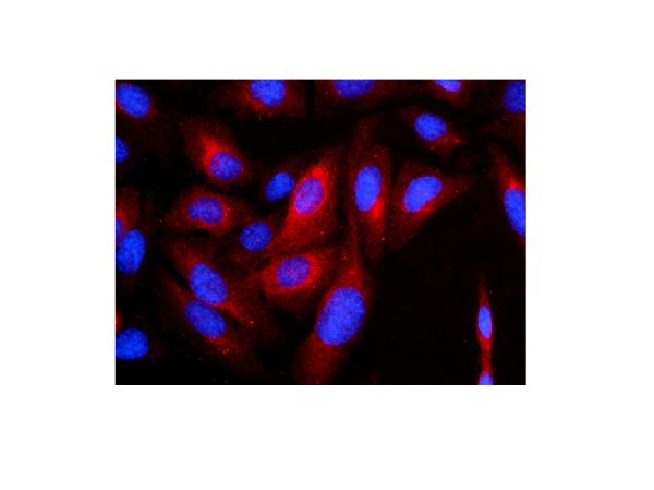
Anti-Cytokeratin 8 KRT8 Antibody Picoband™ (monoclonal, 3G9)
- SPECIFICATION
- CITATIONS
- PROTOCOLS
- BACKGROUND

Application
| WB, IHC, IHC-F, IF, ICC, FC |
|---|---|
| Primary Accession | P05787 |
| Host | Mouse |
| Isotype | Mouse IgG2b |
| Reactivity | Rat, Human, Mouse |
| Clonality | Monoclonal |
| Format | Lyophilized |
| Description | Anti-Cytokeratin 8 KRT8 Antibody Picoband™ (monoclonal, 3G9) . Tested in Flow Cytometry, IF, IHC, IHC-F, ICC, WB applications. This antibody reacts with Human, Mouse, Rat. |
| Reconstitution | Add 0.2ml of distilled water will yield a concentration of 500 µg/ml. |
| Gene ID | 3856 |
|---|---|
| Other Names | Keratin, type II cytoskeletal 8, Cytokeratin-8, CK-8, Keratin-8, K8, Type-II keratin Kb8, KRT8, CYK8 |
| Calculated MW | 54 kDa |
| Application Details | Western blot, 0.1-0.5 µg/ml Immunohistochemistry (Paraffin-embedded Section), 0.5-1 µg/ml Immunohistochemistry (Frozen Section), 0.5-1 µg/ml Immunofluorescence, 2 µg/ml Immunocytochemistry/Immunofluorescence, 2 µg/ml Flow Cytometry, 1-3 µg/1x10^6 cells |
| Subcellular Localization | Nucleoplasm. Nucleus matrix. Cytoplasm. |
| Tissue Specificity | Observed in muscle fibers accumulating in the costameres of myoplasm at the sarcolemma membrane in structures that contain dystrophin and spectrin. Expressed in gingival mucosa and hard palate of the oral cavity. |
| Contents | Each vial contains 4mg Trehalose, 0.9mg NaCl, 0.2mg Na2HPO4, 0.05mg NaN3. |
| Clone Names | Clone: 3G9 |
| Immunogen | E.coli-derived human Cytokeratin 8 recombinant protein (Position: D107-K325). Human Cytokeratin 8 shares 95.4% and 94.5% amino acid (aa) sequence identity with mouse and rat Cytokeratin 8, respectively. |
| Cross Reactivity | No cross-reactivity with other proteins. |
| Storage | Store at -20˚C for one year from date of receipt. After reconstitution, at 4˚C for one month. It can also be aliquotted and stored frozen at -20˚C for six months. Avoid repeated freeze-thaw cycles. |
| Name | KRT8 |
|---|---|
| Synonyms | CYK8 |
| Function | Together with KRT19, helps to link the contractile apparatus to dystrophin at the costameres of striated muscle. |
| Cellular Location | Cytoplasm. Nucleus, nucleoplasm {ECO:0000250|UniProtKB:Q10758}. Nucleus matrix {ECO:0000250|UniProtKB:Q10758} |
| Tissue Location | Observed in muscle fibers accumulating in the costameres of myoplasm at the sarcolemma membrane in structures that contain dystrophin and spectrin. Expressed in gingival mucosa and hard palate of the oral cavity. |

Thousands of laboratories across the world have published research that depended on the performance of antibodies from Abcepta to advance their research. Check out links to articles that cite our products in major peer-reviewed journals, organized by research category.
info@abcepta.com, and receive a free "I Love Antibodies" mug.

Provided below are standard protocols that you may find useful for product applications.
Background
Keratin, type II cytoskeletal 8, also known as cytokeratin-8 (CK-8) or keratin-8 (K8) is a keratin protein that is encoded in humans by the KRT8 gene. This gene is a member of the type II keratin family clustered on the long arm of chromosome 12. Type I and type II keratins heteropolymerize to form intermediate-sized filaments in the cytoplasm of epithelial cells. The product of this gene typically dimerizes with keratin 18 to form an intermediate filament in simple single-layered epithelial cells. This protein plays a role in maintaining cellular structural integrity and also functions in signal transduction and cellular differentiation. Mutations in this gene cause cryptogenic cirrhosis. Alternatively spliced transcript variants have been found for this gene.
If you have used an Abcepta product and would like to share how it has performed, please click on the "Submit Review" button and provide the requested information. Our staff will examine and post your review and contact you if needed.
If you have any additional inquiries please email technical services at tech@abcepta.com.
Ordering Information
Other Products
Shipping Information
