


 Foundational characteristics of cancer include proliferation, angiogenesis, migration, evasion of apoptosis, and cellular immortality. Find key markers for these cellular processes and antibodies to detect them.
 The SUMOplot™ Analysis Program predicts and scores sumoylation sites in your protein. SUMOylation is a post-translational modification involved in various cellular processes, such as nuclear-cytosolic transport, transcriptional regulation, apoptosis, protein stability, response to stress, and progression through the cell cycle.
 The Autophagy Receptor Motif Plotter predicts and scores autophagy receptor binding sites in your protein. Identifying proteins connected to this pathway is critical to understanding the role of autophagy in physiological as well as pathological processes such as development, differentiation, neurodegenerative diseases, stress, infection, and cancer.
Anti-Cystatin C/CST3 Antibody Picoband™ (monoclonal, 4H8)
- SPECIFICATION
- CITATIONS
- PROTOCOLS
- BACKGROUND

Application
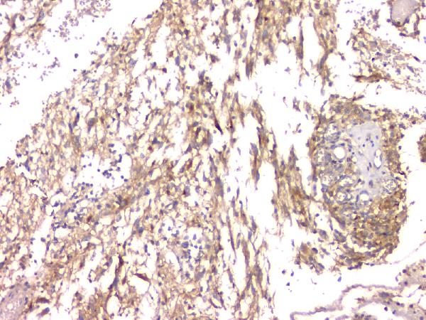
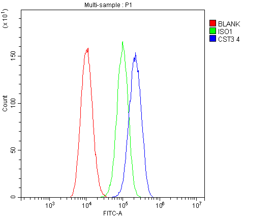
| WB, IHC, IF, ICC, FC, E |
|---|---|
| Primary Accession | P01034 |
| Host | Mouse |
| Isotype | Mouse IgG2a |
| Reactivity | Human |
| Clonality | Monoclonal |
| Format | Lyophilized |
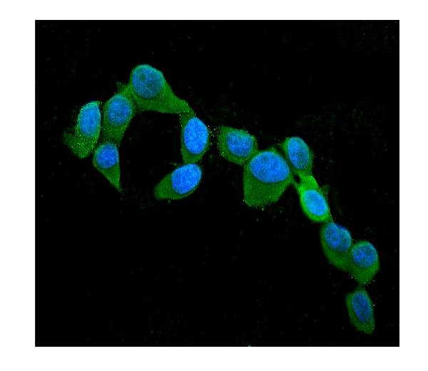
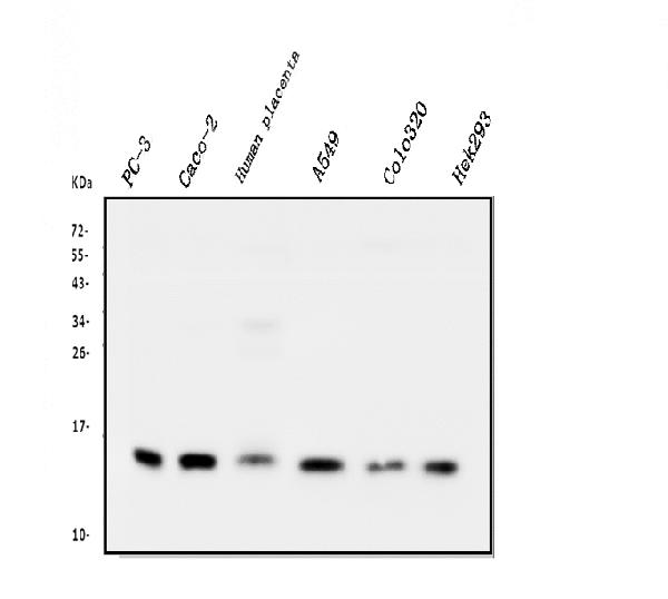
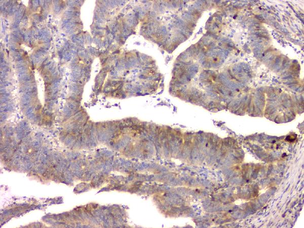
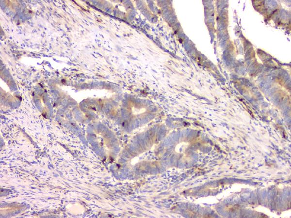
| Description | Anti-Cystatin C/CST3 Antibody Picoband™ (monoclonal, 4H8) . Tested in ELISA, Flow Cytometry, IF, IHC, ICC, WB applications. This antibody reacts with Human. |
| Reconstitution | Add 0.2ml of distilled water will yield a concentration of 500 µg/ml. |
| Gene ID | 1471 |
|---|---|
| Other Names | Cystatin-C, Cystatin-3, Gamma-trace, Neuroendocrine basic polypeptide, Post-gamma-globulin, CST3 |
| Calculated MW | 15 kDa |
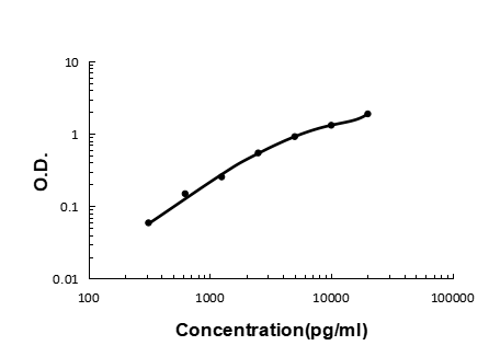
| Application Details | Western blot, 0.1-0.5 µg/ml Immunohistochemistry (Paraffin-embedded Section), 0.5-1 µg/ml Immunocytochemistry/Immunofluorescence, 2 µg/ml Flow Cytometry, 1-3 µg/1x10^6 cells ELISA (Cap), 1-5 µg/ml |
| Subcellular Localization | Nucleus |
| Tissue Specificity | Expressed in submandibular and sublingual saliva but not in parotid saliva (at protein level). Expressed in various body fluids, such as the cerebrospinal fluid and plasma. Expressed in highest levels in the epididymis, vas deferens, brain, thymus, and ovary and the lowest in the submandibular gland. |
| Contents | Each vial contains 4mg Trehalose, 0.9mg NaCl, 0.2mg Na2HPO4, 0.05mg NaN3. |
| Clone Names | Clone: 4H8 |
| Immunogen | E. coli-derived human Cystatin C recombinant protein (Position: K31-A146). |
| Cross Reactivity | No cross-reactivity with other proteins. |
| Storage | Store at -20˚C for one year from date of receipt. After reconstitution, at 4˚C for one month. It can also be aliquotted and stored frozen at -20˚C for six months. Avoid repeated freeze-thaw cycles. |
| Name | CST3 |
|---|---|
| Function | As an inhibitor of cysteine proteinases, this protein is thought to serve an important physiological role as a local regulator of this enzyme activity. |
| Cellular Location | Secreted. |
| Tissue Location | Expressed in submandibular and sublingual saliva but not in parotid saliva (at protein level). Expressed in various body fluids, such as the cerebrospinal fluid and plasma. Expressed in highest levels in the epididymis, vas deferens, brain, thymus, and ovary and the lowest in the submandibular gland |

Thousands of laboratories across the world have published research that depended on the performance of antibodies from Abcepta to advance their research. Check out links to articles that cite our products in major peer-reviewed journals, organized by research category.
info@abcepta.com, and receive a free "I Love Antibodies" mug.

Provided below are standard protocols that you may find useful for product applications.
Background
Cystatin C or cystatin 3, a protein encoded by the CST3 gene, is mainly used as a biomarker of kidney function. Recently, it has been studied for its role in predicting new-onset or deteriorating cardiovascular disease. It also seems to play a role in brain disorders involving amyloid, such as Alzheimer's disease. In humans, all cells with a nucleus (cell core containing the DNA) produce cystatin C as a chain of 120 amino acids. It is found in virtually all tissues and body fluids. It is a potent inhibitor of lysosomal proteinases (enzymes from a special subunit of the cell that break down proteins) and probably one of the most important extracellular inhibitors of cysteine proteases (it prevents the breakdown of proteins outside the cell by a specific type of protein degrading enzymes). Cystatin C belongs to the type 2 cystatin gene family.
If you have used an Abcepta product and would like to share how it has performed, please click on the "Submit Review" button and provide the requested information. Our staff will examine and post your review and contact you if needed.
If you have any additional inquiries please email technical services at tech@abcepta.com.
Ordering Information
Other Products
Shipping Information
