


 Foundational characteristics of cancer include proliferation, angiogenesis, migration, evasion of apoptosis, and cellular immortality. Find key markers for these cellular processes and antibodies to detect them.
 The SUMOplot™ Analysis Program predicts and scores sumoylation sites in your protein. SUMOylation is a post-translational modification involved in various cellular processes, such as nuclear-cytosolic transport, transcriptional regulation, apoptosis, protein stability, response to stress, and progression through the cell cycle.
 The Autophagy Receptor Motif Plotter predicts and scores autophagy receptor binding sites in your protein. Identifying proteins connected to this pathway is critical to understanding the role of autophagy in physiological as well as pathological processes such as development, differentiation, neurodegenerative diseases, stress, infection, and cancer.
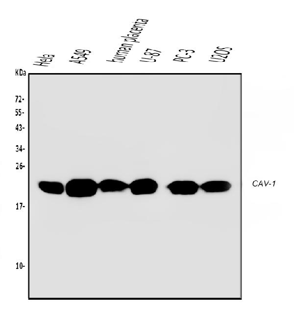
Anti-Caveolin-1/CAV1 Antibody Picoband™ (monoclonal, 12C7)
- SPECIFICATION
- CITATIONS
- PROTOCOLS
- BACKGROUND

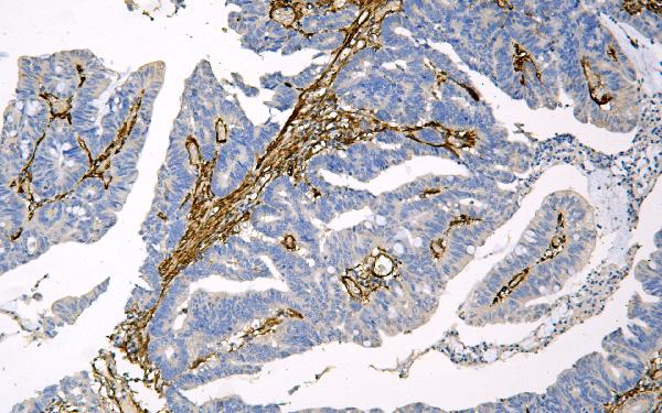
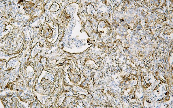
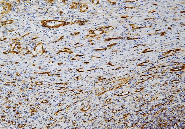
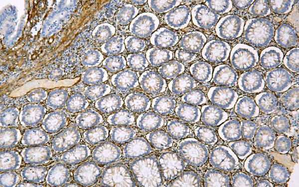
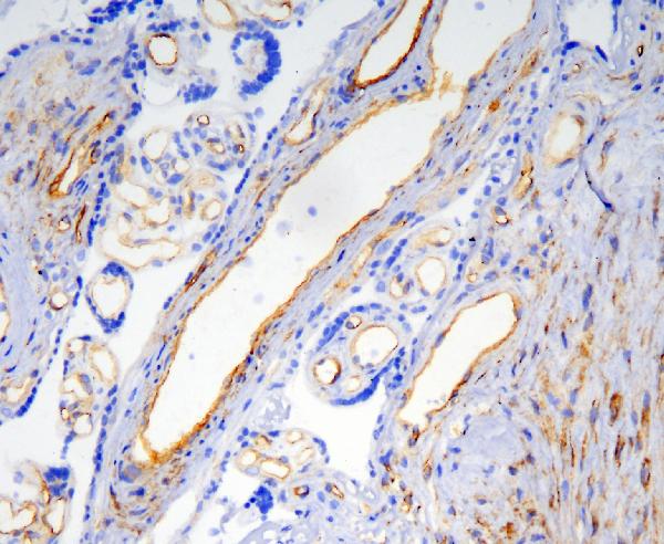
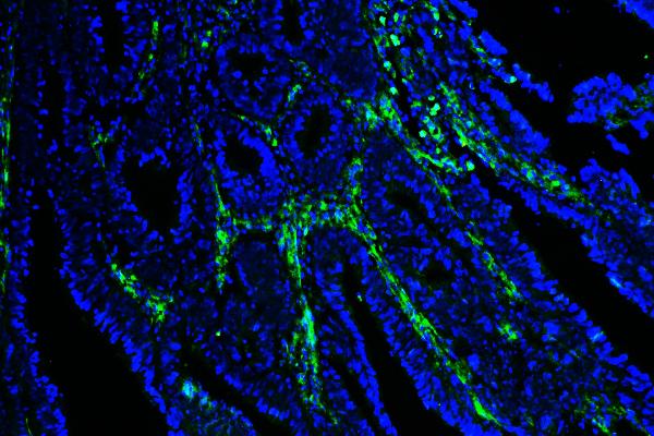
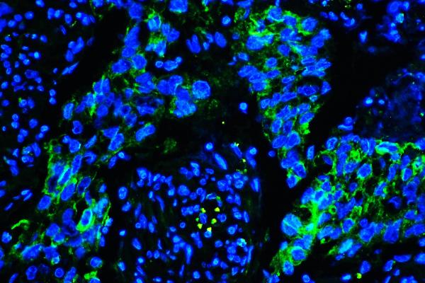
Application
| WB, IHC, IHC-F, IF |
|---|---|
| Primary Accession | Q03135 |
| Host | Mouse |
| Isotype | Mouse IgG2a |
| Reactivity | Human |
| Clonality | Monoclonal |
| Format | Lyophilized |
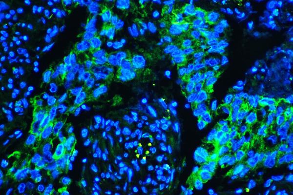
| Description | Anti-Caveolin-1/CAV1 Antibody Picoband™ (monoclonal, 12C7) . Tested in IF, IHC, IHC-F, WB applications. This antibody reacts with Human. |
| Reconstitution | Add 0.2ml of distilled water will yield a concentration of 500 µg/ml. |
| Gene ID | 857 |
|---|---|
| Other Names | Caveolin-1, CAV1, CAV |
| Calculated MW | 22 kDa |
| Application Details | Western blot, 0.1-0.5 µg/ml Immunohistochemistry (Paraffin-embedded Section), 0.5-1 µg/ml Immunohistochemistry (Frozen Section), 0.5-1 µg/ml Immunofluorescence, 2 µg/ml |
| Subcellular Localization | Cell membrane. Peripheral membrane protein. Golgi apparatus membrane. Peripheral membrane protein. Trans-Golgi network. Caveola. Peripheral membrane protein. Membrane raft. |
| Tissue Specificity | Skeletal muscle, liver, stomach, lung, kidney and heart (at protein level). Expressed in the brain. |
| Contents | Each vial contains 4mg Trehalose, 0.9mg NaCl, 0.2mg Na2HPO4, 0.05mg NaN3. |
| Clone Names | Clone: 12C7 |
| Immunogen | E.coli-derived human Caveolin-1 recombinant protein (Position: G4-I178). Human Caveolin-1 shares 95% and 94% amino acid (aa) sequence identity with mouse and rat Caveolin-1, respectively. |
| Cross Reactivity | No cross-reactivity with other proteins. |
| Storage | Store at -20˚C for one year from date of receipt. After reconstitution, at 4˚C for one month. It can also be aliquotted and stored frozen at -20˚C for six months. Avoid repeated freeze-thaw cycles. |
| Name | CAV1 |
|---|---|
| Synonyms | CAV |
| Function | May act as a scaffolding protein within caveolar membranes (PubMed:11751885). Forms a stable heterooligomeric complex with CAV2 that targets to lipid rafts and drives caveolae formation. Mediates the recruitment of CAVIN proteins (CAVIN1/2/3/4) to the caveolae (PubMed:19262564). Interacts directly with G-protein alpha subunits and can functionally regulate their activity (By similarity). Involved in the costimulatory signal essential for T-cell receptor (TCR)-mediated T-cell activation. Its binding to DPP4 induces T-cell proliferation and NF-kappa-B activation in a T-cell receptor/CD3-dependent manner (PubMed:17287217). Recruits CTNNB1 to caveolar membranes and may regulate CTNNB1-mediated signaling through the Wnt pathway (By similarity). Negatively regulates TGFB1-mediated activation of SMAD2/3 by mediating the internalization of TGFBR1 from membrane rafts leading to its subsequent degradation (PubMed:25893292). Binds 20(S)- hydroxycholesterol (20(S)-OHC) (By similarity). |
| Cellular Location | Golgi apparatus membrane; Peripheral membrane protein. Cell membrane; Peripheral membrane protein. Membrane, caveola; Peripheral membrane protein. Membrane raft. Golgi apparatus, trans-Golgi network {ECO:0000250|UniProtKB:P33724} Note=Colocalized with DPP4 in membrane rafts. Potential hairpin-like structure in the membrane. Membrane protein of caveolae |
| Tissue Location | Skeletal muscle, liver, stomach, lung, kidney and heart (at protein level). Expressed in the brain |

Thousands of laboratories across the world have published research that depended on the performance of antibodies from Abcepta to advance their research. Check out links to articles that cite our products in major peer-reviewed journals, organized by research category.
info@abcepta.com, and receive a free "I Love Antibodies" mug.

Provided below are standard protocols that you may find useful for product applications.
Background
CAV1 ( Caveolin-1) is a protein that in humans is encoded by the CAV1 gene. The CAV1 gene is mapped to 7q31.2. The scaffolding protein encoded by this gene is the main component of the caveolae plasma membranes found in most cell types. The protein links integrin subunits to the tyrosine kinase FYN, an initiating step in coupling integrins to the Ras-ERK pathway and promoting cell cycle progression. The gene is a tumor suppressor gene candidate and a negative regulator of the Ras-p42/44 MAP kinase cascade. CAV1 and CAV2 are located next to each other on chromosome 7 and express colocalizing proteins that form a stable hetero-oligomeric complex. By using alternative initiation codons in the same reading frame, two isoforms (alpha and beta) are encoded by a single transcript from this gene.
If you have used an Abcepta product and would like to share how it has performed, please click on the "Submit Review" button and provide the requested information. Our staff will examine and post your review and contact you if needed.
If you have any additional inquiries please email technical services at tech@abcepta.com.
Ordering Information
Other Products
Shipping Information
