


 Foundational characteristics of cancer include proliferation, angiogenesis, migration, evasion of apoptosis, and cellular immortality. Find key markers for these cellular processes and antibodies to detect them.
 The SUMOplot™ Analysis Program predicts and scores sumoylation sites in your protein. SUMOylation is a post-translational modification involved in various cellular processes, such as nuclear-cytosolic transport, transcriptional regulation, apoptosis, protein stability, response to stress, and progression through the cell cycle.
 The Autophagy Receptor Motif Plotter predicts and scores autophagy receptor binding sites in your protein. Identifying proteins connected to this pathway is critical to understanding the role of autophagy in physiological as well as pathological processes such as development, differentiation, neurodegenerative diseases, stress, infection, and cancer.
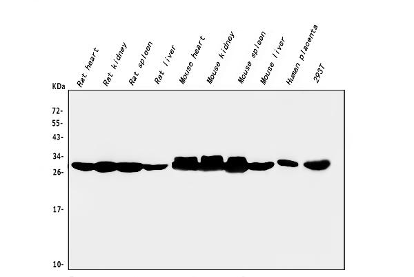
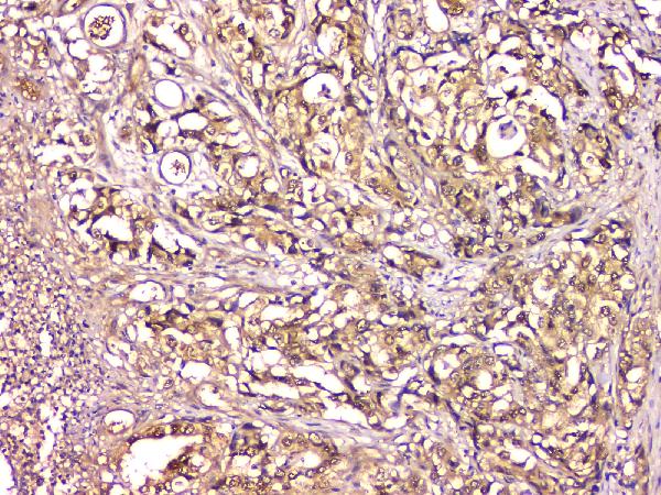
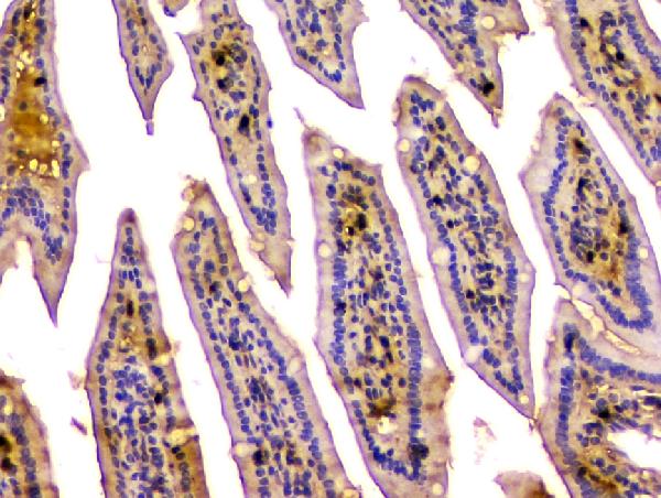
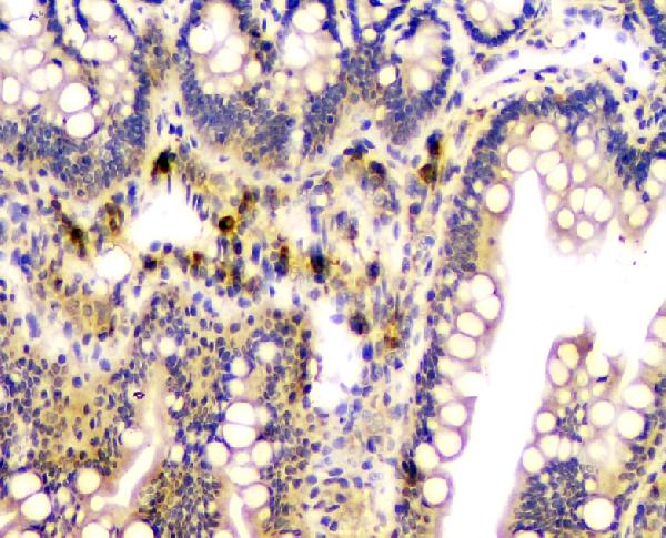
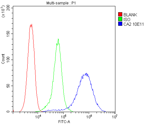
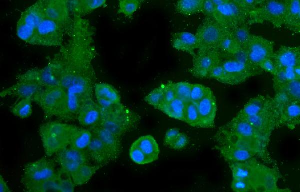
Anti-CA2 Antibody Picoband™ (monoclonal, 10E11)

- SPECIFICATION
- CITATIONS
- PROTOCOLS
- BACKGROUND

Application
| WB, IHC, IF, ICC, FC |
|---|---|
| Primary Accession | P00918 |
| Host | Mouse |
| Isotype | Mouse IgG2b |
| Reactivity | Rat, Human, Mouse |
| Clonality | Monoclonal |
| Format | Lyophilized |
| Description | Anti-CA2 Antibody Picoband™ (monoclonal, 10E11) . Tested in Flow Cytometry, IF, IHC, ICC, WB applications. This antibody reacts with Human, Mouse, Rat. |
| Reconstitution | Add 0.2ml of distilled water will yield a concentration of 500 µg/ml. |
| Gene ID | 760 |
|---|---|
| Other Names | Carbonic anhydrase 2, 4.2.1.1, Carbonate dehydratase II, Carbonic anhydrase C, CAC, Carbonic anhydrase II, CA-II, Cyanamide hydratase CA2, 4.2.1.69, CA2 |
| Calculated MW | 28 kDa |
| Application Details | Western blot, 0.1-0.5 µg/ml Immunohistochemistry (Paraffin-embedded Section), 0.5-1 µg/ml Immunocytochemistry/Immunofluorescence, 5 µg/ml Flow Cytometry, 1-3 µg/1x10^6 cells |
| Subcellular Localization | Cell membrane. Cytoplasm. |
| Contents | Each vial contains 4mg Trehalose, 0.9mg NaCl, 0.2mg Na2HPO4, 0.05mg NaN3. |
| Clone Names | Clone: 10E11 |
| Immunogen | E.coli-derived human CA2 recombinant protein (Position: S2-K260). Human CA2 shares 81.1% and 80.7% amino acid (aa) sequence identity with mouse and rat CA2, respectively. |
| Cross Reactivity | No cross-reactivity with other proteins. |
| Storage | Store at -20˚C for one year from date of receipt. After reconstitution, at 4˚C for one month. It can also be aliquotted and stored frozen at -20˚C for six months. Avoid repeated freeze-thaw cycles. |
| Name | CA2 |
|---|---|
| Function | Catalyzes the reversible hydration of carbon dioxide (PubMed:11327835, PubMed:11802772, PubMed:11831900, PubMed:12056894, PubMed:12171926, PubMed:1336460, PubMed:14736236, PubMed:15300855, PubMed:15453828, PubMed:15667203, PubMed:15865431, PubMed:16106378, PubMed:16214338, PubMed:16290146, PubMed:16686544, PubMed:16759856, PubMed:16807956, PubMed:17127057, PubMed:17251017, PubMed:17314045, PubMed:17330962, PubMed:17346964, PubMed:17540563, PubMed:17588751, PubMed:17705204, PubMed:18024029, PubMed:18162396, PubMed:18266323, PubMed:18374572, PubMed:18481843, PubMed:18618712, PubMed:18640037, PubMed:18942852, PubMed:1909891, PubMed:1910042, PubMed:19170619, PubMed:19186056, PubMed:19206230, PubMed:19520834, PubMed:19778001, PubMed:7761440, PubMed:7901850, PubMed:8218160, PubMed:8262987, PubMed:8399159, PubMed:8451242, PubMed:8485129, PubMed:8639494, PubMed:9265618, PubMed:9398308). Can also hydrate cyanamide to urea (PubMed:10550681, PubMed:11015219). Stimulates the chloride-bicarbonate exchange activity of SLC26A6 (PubMed:15990874). Essential for bone resorption and osteoclast differentiation (PubMed:15300855). Involved in the regulation of fluid secretion into the anterior chamber of the eye. Contributes to intracellular pH regulation in the duodenal upper villous epithelium during proton-coupled peptide absorption. |
| Cellular Location | Cytoplasm. Cell membrane. Note=Colocalized with SLC26A6 at the surface of the cell membrane in order to form a bicarbonate transport metabolon. Displaced from the cytosolic surface of the cell membrane by PKC in phorbol myristate acetate (PMA)-induced cells |

Thousands of laboratories across the world have published research that depended on the performance of antibodies from Abcepta to advance their research. Check out links to articles that cite our products in major peer-reviewed journals, organized by research category.
info@abcepta.com, and receive a free "I Love Antibodies" mug.

Provided below are standard protocols that you may find useful for product applications.
Background
CA2 is a cytosolic enzyme with the highest activity among all known CAs. The carbonic anhydrases (ACs) form a family of enzymes that catalyze the rapid interconversion of carbon dioxide and water to bicarbonate and protons (or vice versa), a reversible reaction that occurs relatively slowly in the absence of a catalyst. Mutations in the CA2 gene result in the CA II deficiency syndrome, an autosomal recessive disorder that produces osteopetrosis, renal tubular acidosis and cerebral calcification. This gene is mapped to 8q22.
If you have used an Abcepta product and would like to share how it has performed, please click on the "Submit Review" button and provide the requested information. Our staff will examine and post your review and contact you if needed.
If you have any additional inquiries please email technical services at tech@abcepta.com.
Ordering Information
Other Products
Shipping Information
