


 Foundational characteristics of cancer include proliferation, angiogenesis, migration, evasion of apoptosis, and cellular immortality. Find key markers for these cellular processes and antibodies to detect them.
 The SUMOplot™ Analysis Program predicts and scores sumoylation sites in your protein. SUMOylation is a post-translational modification involved in various cellular processes, such as nuclear-cytosolic transport, transcriptional regulation, apoptosis, protein stability, response to stress, and progression through the cell cycle.
 The Autophagy Receptor Motif Plotter predicts and scores autophagy receptor binding sites in your protein. Identifying proteins connected to this pathway is critical to understanding the role of autophagy in physiological as well as pathological processes such as development, differentiation, neurodegenerative diseases, stress, infection, and cancer.
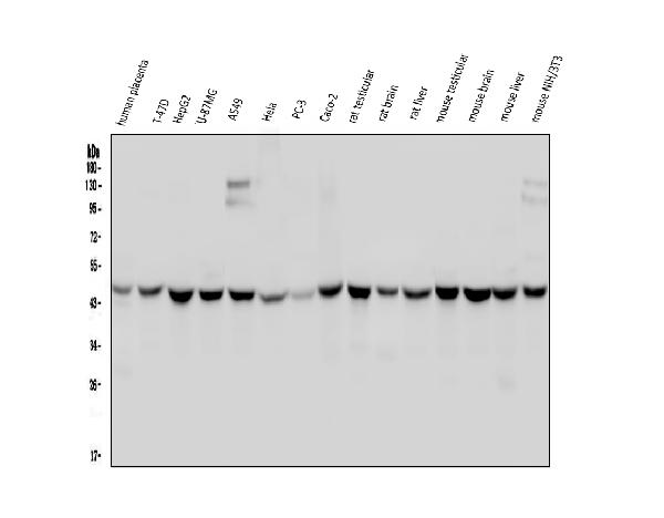
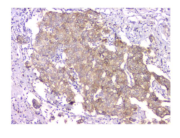
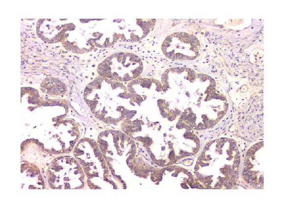
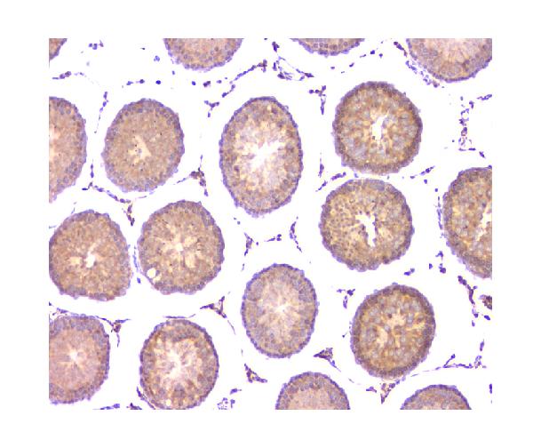
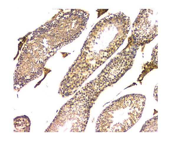
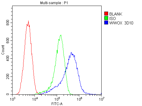
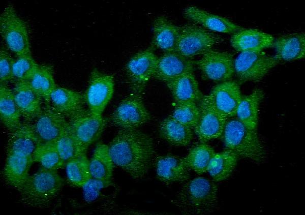
Anti-WWOX Antibody Picoband™ (monoclonal, 3D10)
- SPECIFICATION
- CITATIONS
- PROTOCOLS
- BACKGROUND

Application
| WB, IHC, IF, ICC, FC |
|---|---|
| Primary Accession | Q9NZC7 |
| Host | Mouse |
| Isotype | Mouse IgG1 |
| Reactivity | Rat, Human, Mouse |
| Clonality | Monoclonal |
| Format | Lyophilized |
| Description | Anti-WWOX Antibody Picoband™ (monoclonal, 3D10) . Tested in Flow Cytometry, IF, IHC, ICC, WB applications. This antibody reacts with Human, Mouse, Rat. |
| Reconstitution | Add 0.2ml of distilled water will yield a concentration of 500 µg/ml. |
| Gene ID | 51741 |
|---|---|
| Other Names | WW domain-containing oxidoreductase, 1.1.1.-, Fragile site FRA16D oxidoreductase, Short chain dehydrogenase/reductase family 41C member 1, WWOX, FOR, SDR41C1, WOX1 |
| Calculated MW | 47 kDa |
| Application Details | Western blot, 0.1-0.5 µg/ml Immunohistochemistry (Paraffin-embedded Section), 0.5-1 µg/ml Immunocytochemistry/Immunofluorescence, 5 µg/ml, Human Flow Cytometry, 1-3 µg/1x10^6 cells |
| Subcellular Localization | Mitochondrion. Golgi apparatus. Nucleus. Cytoplasm. |
| Tissue Specificity | Widely expressed. Strongly expressed in testis, prostate, and ovary. Overexpressed in cancer cell lines. Isoform 5 and isoform 6 may only be expressed in tumor cell lines. |
| Contents | Each vial contains 4mg Trehalose, 0.9mg NaCl, 0.2mg Na2HPO4, 0.05mg NaN3. |
| Clone Names | Clone: 3D10 |
| Immunogen | E. coli-derived human WWOX recombinant protein (Position: M1-D245). |
| Cross Reactivity | No cross-reactivity with other proteins. |
| Storage | Store at -20˚C for one year from date of receipt. After reconstitution, at 4˚C for one month. It can also be aliquotted and stored frozen at -20˚C for six months. Avoid repeated freeze-thaw cycles. |
| Name | WWOX |
|---|---|
| Synonyms | FOR, SDR41C1, WOX1 |
| Function | Putative oxidoreductase. Acts as a tumor suppressor and plays a role in apoptosis. Required for normal bone development (By similarity). May function synergistically with p53/TP53 to control genotoxic stress-induced cell death. Plays a role in TGFB1 signaling and TGFB1-mediated cell death. May also play a role in tumor necrosis factor (TNF)-mediated cell death. Inhibits Wnt signaling, probably by sequestering DVL2 in the cytoplasm. |
| Cellular Location | Cytoplasm. Nucleus Mitochondrion. Golgi apparatus. Lysosome Note=Partially localizes to the mitochondria (PubMed:14695174) Translocates to the nucleus upon genotoxic stress or TNF stimulation (By similarity). Translocates to the nucleus in response to TGFB1 (PubMed:19366691). Isoform 5 and isoform 6 may localize in the nucleus Localized to the lysosome probably upon binding to VOPP1 (PubMed:30285739). {ECO:0000250, ECO:0000269|PubMed:14695174, ECO:0000269|PubMed:19366691, ECO:0000269|PubMed:30285739} |
| Tissue Location | Widely expressed. Strongly expressed in testis, prostate, and ovary. Overexpressed in cancer cell lines. Isoform 5 and isoform 6 may only be expressed in tumor cell lines |

Thousands of laboratories across the world have published research that depended on the performance of antibodies from Abcepta to advance their research. Check out links to articles that cite our products in major peer-reviewed journals, organized by research category.
info@abcepta.com, and receive a free "I Love Antibodies" mug.

Provided below are standard protocols that you may find useful for product applications.
Background
WW domain-containing oxidoreductase is an enzyme that in humans is encoded by the WWOX gene. This gene encodes a member of the short-chain dehydrogenases/reductases (SDR) protein family. It spans the FRA16D common chromosomal fragile site and appears to function as a tumor suppressor gene. Expression of the encoded protein is able to induce apoptosis, while defects in this gene are associated with multiple types of cancer. Disruption of this gene is also associated with autosomal recessive spinocerebellar ataxia 12. Disruption of a similar gene in mouse results in impaired steroidogenesis, additionally suggesting a metabolic function for the protein. Alternative splicing results in multiple transcript variants.
If you have used an Abcepta product and would like to share how it has performed, please click on the "Submit Review" button and provide the requested information. Our staff will examine and post your review and contact you if needed.
If you have any additional inquiries please email technical services at tech@abcepta.com.
Ordering Information
Other Products
Shipping Information
