


 Foundational characteristics of cancer include proliferation, angiogenesis, migration, evasion of apoptosis, and cellular immortality. Find key markers for these cellular processes and antibodies to detect them.
 The SUMOplot™ Analysis Program predicts and scores sumoylation sites in your protein. SUMOylation is a post-translational modification involved in various cellular processes, such as nuclear-cytosolic transport, transcriptional regulation, apoptosis, protein stability, response to stress, and progression through the cell cycle.
 The Autophagy Receptor Motif Plotter predicts and scores autophagy receptor binding sites in your protein. Identifying proteins connected to this pathway is critical to understanding the role of autophagy in physiological as well as pathological processes such as development, differentiation, neurodegenerative diseases, stress, infection, and cancer.
> home > Products > Primary Antibodies > Signal Transduction > Anti-SHP2/PTPN11 Antibody Picoband™ (monoclonal, 2E6)
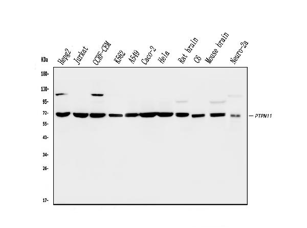
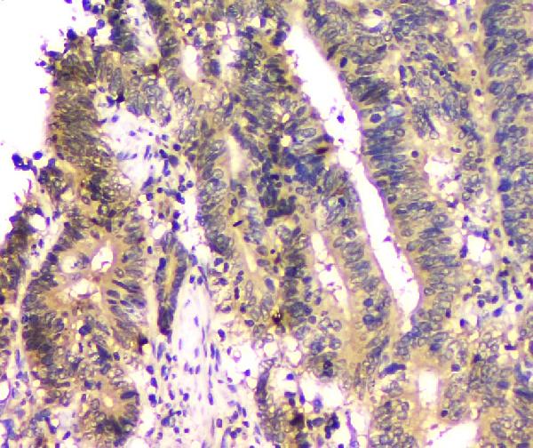
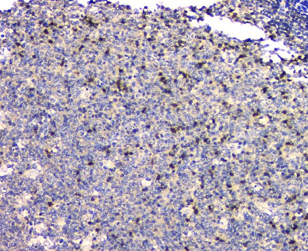
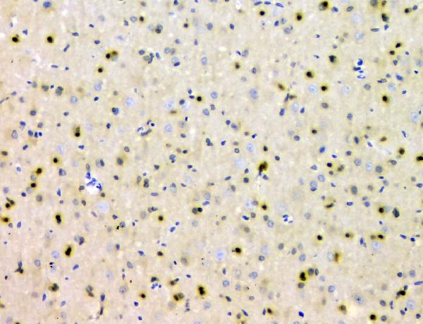
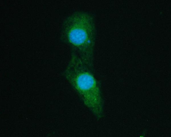
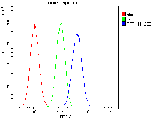
Anti-SHP2/PTPN11 Antibody Picoband™ (monoclonal, 2E6)

- SPECIFICATION
- CITATIONS
- PROTOCOLS
- BACKGROUND

Application
| WB, IHC, IF, ICC, FC |
|---|---|
| Primary Accession | Q06124 |
| Host | Mouse |
| Isotype | Mouse IgG2b |
| Reactivity | Rat, Human, Mouse |
| Clonality | Monoclonal |
| Format | Lyophilized |
| Description | Anti-SHP2/PTPN11 Antibody Picoband™ (monoclonal, 2E6) . Tested in Flow Cytometry, IF, IHC, ICC, WB applications. This antibody reacts with Human, Mouse, Rat. |
| Reconstitution | Add 0.2ml of distilled water will yield a concentration of 500 µg/ml. |
| Gene ID | 5781 |
|---|---|
| Other Names | Tyrosine-protein phosphatase non-receptor type 11, 3.1.3.48, Protein-tyrosine phosphatase 1D, PTP-1D, Protein-tyrosine phosphatase 2C, PTP-2C, SH-PTP2, SHP-2, Shp2, SH-PTP3, PTPN11, PTP2C, SHPTP2 |
| Calculated MW | 70 kDa |
| Application Details | Western blot, 0.1-0.5 µg/ml Immunohistochemistry (Paraffin-embedded Section), 0.5-1 µg/ml Immunocytochemistry/Immunofluorescence, 2 µg/ml Flow Cytometry, 1-3 µg/1x10^6 cells |
| Subcellular Localization | Nucleus. Cytoplasm. |
| Tissue Specificity | Widely expressed, with highest levels in heart, brain, and skeletal muscle. |
| Contents | Each vial contains 4mg Trehalose, 0.9mg NaCl, 0.2mg Na2HPO4, 0.05mg NaN3. |
| Clone Names | Clone: Clone: 2E6 |
| Immunogen | A synthetic peptide corresponding to a sequence at the N-terminus of human SHP2, identical to the related mouse and rat sequences. |
| Cross Reactivity | No cross-reactivity with other proteins. |
| Storage | Store at -20˚C for one year from date of receipt. After reconstitution, at 4˚C for one month. It can also be aliquotted and stored frozen at -20˚C for six months. Avoid repeated freeze-thaw cycles. |
| Name | PTPN11 |
|---|---|
| Synonyms | PTP2C, SHPTP2 |
| Function | Acts downstream of various receptor and cytoplasmic protein tyrosine kinases to participate in the signal transduction from the cell surface to the nucleus (PubMed:10655584, PubMed:14739280, PubMed:18559669, PubMed:18829466, PubMed:26742426, PubMed:28074573). Positively regulates MAPK signal transduction pathway (PubMed:28074573). Dephosphorylates GAB1, ARHGAP35 and EGFR (PubMed:28074573). Dephosphorylates ROCK2 at 'Tyr-722' resulting in stimulation of its RhoA binding activity (PubMed:18559669). Dephosphorylates CDC73 (PubMed:26742426). Dephosphorylates SOX9 on tyrosine residues, leading to inactivate SOX9 and promote ossification (By similarity). Dephosphorylates tyrosine-phosphorylated NEDD9/CAS-L (PubMed:19275884). |
| Cellular Location | Cytoplasm. Nucleus |
| Tissue Location | Widely expressed, with highest levels in heart, brain, and skeletal muscle. |
Research Areas

Abcepta welcomes feedback from its customers.
If you have used an Abcepta product and would like to share how it has performed, please click on the "Submit Review" button and provide the requested information. Our staff will examine and post your review and contact you if needed.
If you have any additional inquiries please email technical services at tech@abcepta.com.
$ 370.00
Cat# ABO14824
Ordering Information
United States
AlbaniaAustraliaAustriaBelgiumBosnia & HerzegovinaBrazilBulgariaCanadaCentral AmericaChinaCroatiaCyprusCzech RepublicDenmarkEstoniaFinlandFranceGermanyGreeceHong KongHungaryIcelandIndiaIndonesiaIrelandIsraelItalyJapanLatviaLithuaniaLuxembourgMacedoniaMalaysiaMaltaNetherlandsNew ZealandNorwayPakistanPolandPortugalRomaniaSerbiaSingaporeSlovakiaSloveniaSouth AfricaSouth KoreaSpainSwedenSwitzerlandTaiwanTurkeyUnited KingdomUnited StatesVietnamWorldwideOthersMexico
USA Headquarters
(888) 735-7227 / (858) 622-0099 or (858) 875-1900
Other Products
Shipping Information
Domestic orders (in stock items)
Shipped out the same day. Orders placed after 1 PM (PST) will ship out the next business day.
International orders
Contact your local distributors
