


 Foundational characteristics of cancer include proliferation, angiogenesis, migration, evasion of apoptosis, and cellular immortality. Find key markers for these cellular processes and antibodies to detect them.
 The SUMOplot™ Analysis Program predicts and scores sumoylation sites in your protein. SUMOylation is a post-translational modification involved in various cellular processes, such as nuclear-cytosolic transport, transcriptional regulation, apoptosis, protein stability, response to stress, and progression through the cell cycle.
 The Autophagy Receptor Motif Plotter predicts and scores autophagy receptor binding sites in your protein. Identifying proteins connected to this pathway is critical to understanding the role of autophagy in physiological as well as pathological processes such as development, differentiation, neurodegenerative diseases, stress, infection, and cancer.
Anti-AKT2 Antibody Picoband™ (monoclonal, 10C6)
- SPECIFICATION
- CITATIONS
- PROTOCOLS
- BACKGROUND

Application
| WB, IF, ICC, FC |
|---|---|
| Primary Accession | P31751 |
| Host | Mouse |
| Isotype | Mouse IgG1 |
| Reactivity | Human |
| Clonality | Monoclonal |
| Format | Lyophilized |
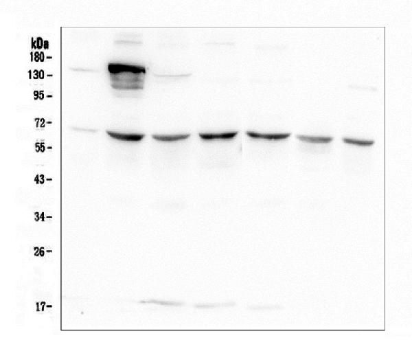
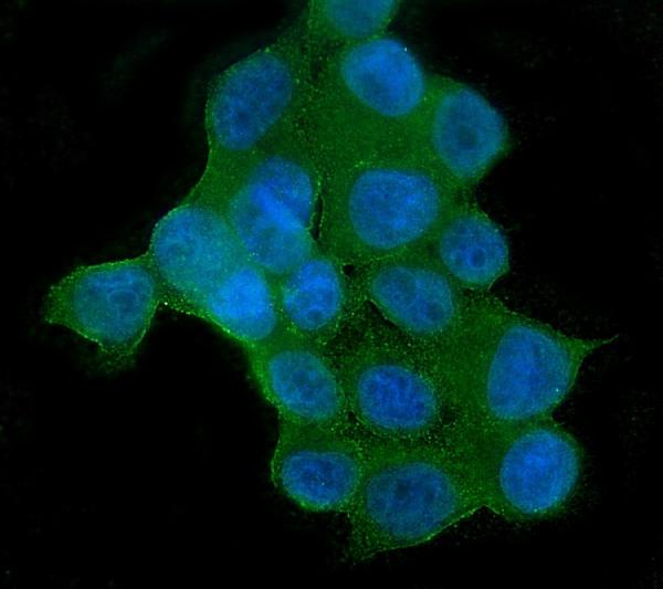
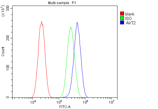
| Description | Anti-AKT2 Antibody Picoband™ (monoclonal, 10C6) . Tested in Flow Cytometry, IF, ICC, WB applications. This antibody reacts with Human. |
| Gene ID | 208 |
|---|---|
| Other Names | RAC-beta serine/threonine-protein kinase, 2.7.11.1, Protein kinase Akt-2, Protein kinase B beta, PKB beta, RAC protein kinase beta, RAC-PK-beta, AKT2 (HGNC:392) |
| Calculated MW | 56 kDa |
| Application Details | Western blot, 0.1-0.5 µg/ml Immunocytochemistry/Immunofluorescence, 2 µg/ml Flow Cytometry, 1-3 µg/1x10^6 cells |
| Subcellular Localization | Cytoplasm. Nucleus. Cell membrane; Peripheral membrane protein. Early endosome |
| Tissue Specificity | Expressed in all cell types so far analyzed. |
| Contents | Each vial contains 4mg Trehalose, 0.9mg NaCl, 0.2mg Na2HPO4, 0.05mg NaN3. |
| Clone Names | Clone: 10C6 |
| Immunogen | A synthetic peptide corresponding to a sequence at the C-terminus of human AKT2, different from the related mouse sequence by two amino acids, and from the related rat sequence by one amino acid. |
| Cross Reactivity | No cross-reactivity with other proteins. |
| Storage | Store at -20˚C for one year from date of receipt. After reconstitution, at 4˚C for one month. It can also be aliquotted and stored frozen at -20˚C for six months. Avoid repeated freeze-thaw cycles. |
| Name | AKT2 (HGNC:392) |
|---|---|
| Function | Serine/threonine kinase closely related to AKT1 and AKT3. All 3 enzymes, AKT1, AKT2 and AKT3, are collectively known as AKT kinase. AKT regulates many processes including metabolism, proliferation, cell survival, growth and angiogenesis, through the phosphorylation of a range of downstream substrates. Over 100 substrates have been reported so far, although for most of them, the precise AKT kinase catalyzing the reaction was not specified. AKT regulates glucose uptake by mediating insulin-induced translocation of the SLC2A4/GLUT4 glucose transporter to the cell surface. Phosphorylation of PTPN1 at 'Ser-50' negatively modulates its phosphatase activity preventing dephosphorylation of the insulin receptor and the attenuation of insulin signaling. Phosphorylation of TBC1D4 triggers the binding of this effector to inhibitory 14-3-3 proteins, which is required for insulin-stimulated glucose transport. AKT also regulates the storage of glucose in the form of glycogen by phosphorylating GSK3A at 'Ser-21' and GSK3B at 'Ser-9', resulting in inhibition of its kinase activity. Phosphorylation of GSK3 isoforms by AKT is also thought to be one mechanism by which cell proliferation is driven. AKT regulates also cell survival via the phosphorylation of MAP3K5 (apoptosis signal- related kinase). Phosphorylation of 'Ser-83' decreases MAP3K5 kinase activity stimulated by oxidative stress and thereby prevents apoptosis. AKT mediates insulin-stimulated protein synthesis by phosphorylating TSC2 at 'Ser-939' and 'Thr-1462', thereby activating mTORC1 signaling and leading to both phosphorylation of 4E-BP1 and in activation of RPS6KB1. AKT is involved in the phosphorylation of members of the FOXO factors (Forkhead family of transcription factors), leading to binding of 14-3-3 proteins and cytoplasmic localization. In particular, FOXO1 is phosphorylated at 'Thr-24', 'Ser-256' and 'Ser-319'. FOXO3 and FOXO4 are phosphorylated on equivalent sites. AKT has an important role in the regulation of NF-kappa-B-dependent gene transcription and positively regulates the activity of CREB1 (cyclic AMP (cAMP)-response element binding protein). The phosphorylation of CREB1 induces the binding of accessory proteins that are necessary for the transcription of pro-survival genes such as BCL2 and MCL1. AKT phosphorylates 'Ser- 454' on ATP citrate lyase (ACLY), thereby potentially regulating ACLY activity and fatty acid synthesis. Activates the 3B isoform of cyclic nucleotide phosphodiesterase (PDE3B) via phosphorylation of 'Ser-273', resulting in reduced cyclic AMP levels and inhibition of lipolysis. Phosphorylates PIKFYVE on 'Ser-318', which results in increased PI(3)P- 5 activity. The Rho GTPase-activating protein DLC1 is another substrate and its phosphorylation is implicated in the regulation cell proliferation and cell growth. AKT plays a role as key modulator of the AKT-mTOR signaling pathway controlling the tempo of the process of newborn neurons integration during adult neurogenesis, including correct neuron positioning, dendritic development and synapse formation. Signals downstream of phosphatidylinositol 3-kinase (PI(3)K) to mediate the effects of various growth factors such as platelet- derived growth factor (PDGF), epidermal growth factor (EGF), insulin and insulin-like growth factor 1 (IGF1). AKT mediates the antiapoptotic effects of IGF1. Essential for the SPATA13-mediated regulation of cell migration and adhesion assembly and disassembly. May be involved in the regulation of the placental development (PubMed:21432781, PubMed:21620960). In response to lysophosphatidic acid stimulation, inhibits the ciliogenesis cascade. In this context, phosphorylates WDR44, hence stabilizing its interaction with Rab11 and preventing the formation of the ciliogenic Rab11-FIP3-RAB3IP complex. Also phosphorylates RAB3IP/Rabin8, thus may affect RAB3IP guanine nucleotide exchange factor (GEF) activity toward Rab8, which is important for cilia growth (PubMed:31204173). Phosphorylates PKP1, facilitating its interaction with YWHAG and translocation to the nucleus, ultimately resulting in a reduction in keratinocyte intercellular adhesion (By similarity). Phosphorylation of PKP1 increases PKP1 protein stability, translocation to the cytoplasm away from desmosome plaques and PKP1- driven cap-dependent translation (PubMed:23444369). |
| Cellular Location | Cytoplasm. Nucleus Cell membrane; Peripheral membrane protein. Early endosome {ECO:0000250|UniProtKB:Q60823}. Note=Through binding of the N-terminal PH domain to phosphatidylinositol (3,4,5)-trisphosphate (PtdIns(3,4,5)P3) or phosphatidylinositol (3,4)-bisphosphate (PtdIns(3,4)P2), recruited to the plasma membrane. Cell membrane recruitment is facilitated by interaction with CLIP3. Colocalizes with WDFY2 in early endosomes (By similarity). Localizes within both nucleus and cytoplasm in proliferative primary myoblasts and mostly within the nucleus of differentiated primary myoblasts (PubMed:17565718) {ECO:0000250|UniProtKB:Q60823, ECO:0000269|PubMed:17565718} |
| Tissue Location | Widely expressed. Expressed in myoblasts (PubMed:17565718). |

Thousands of laboratories across the world have published research that depended on the performance of antibodies from Abcepta to advance their research. Check out links to articles that cite our products in major peer-reviewed journals, organized by research category.
info@abcepta.com, and receive a free "I Love Antibodies" mug.

Provided below are standard protocols that you may find useful for product applications.
Background
AKT2 is a putative oncogene encoding a protein belonging to a subfamily of serine/threonine kinases containing SH2-like (Src homology 2-like) domains. This gene is mapped to 19q13.2. AKT2 is one of 3 closely related serine/threonine-protein kinases (AKT1, AKT2 and AKT3) called the AKT kinase, and which regulate many processes including metabolism, proliferation, cell survival, growth and angiogenesis. AKT2 seems also to be the principal isoform responsible of the regulation of glucose uptake. AKT2 is also specifically involved in skeletal muscle differentiation, one of its substrates in this process being ANKRD2. Overexpression of AKT2 contributes to the malignant phenotype of a subset of human ductal pancreatic cancers.
If you have used an Abcepta product and would like to share how it has performed, please click on the "Submit Review" button and provide the requested information. Our staff will examine and post your review and contact you if needed.
If you have any additional inquiries please email technical services at tech@abcepta.com.
Ordering Information
Other Products
Shipping Information
