


 Foundational characteristics of cancer include proliferation, angiogenesis, migration, evasion of apoptosis, and cellular immortality. Find key markers for these cellular processes and antibodies to detect them.
 The SUMOplot™ Analysis Program predicts and scores sumoylation sites in your protein. SUMOylation is a post-translational modification involved in various cellular processes, such as nuclear-cytosolic transport, transcriptional regulation, apoptosis, protein stability, response to stress, and progression through the cell cycle.
 The Autophagy Receptor Motif Plotter predicts and scores autophagy receptor binding sites in your protein. Identifying proteins connected to this pathway is critical to understanding the role of autophagy in physiological as well as pathological processes such as development, differentiation, neurodegenerative diseases, stress, infection, and cancer.
Anti-CD2AP Antibody Picoband™ (monoclonal, 5F8)


- SPECIFICATION
- CITATIONS
- PROTOCOLS
- BACKGROUND

Application
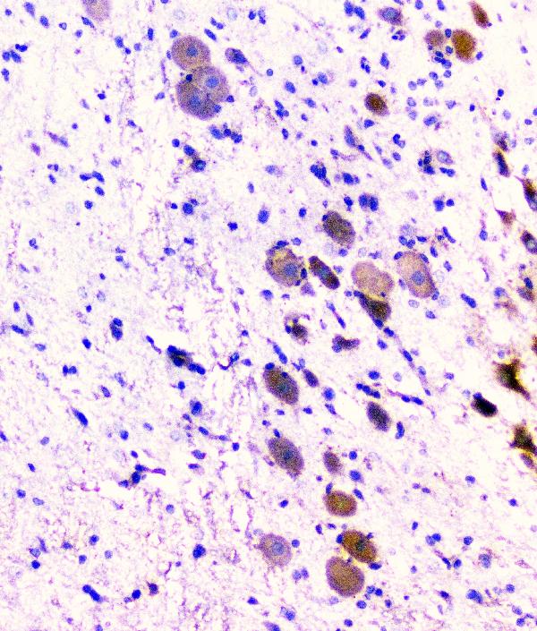
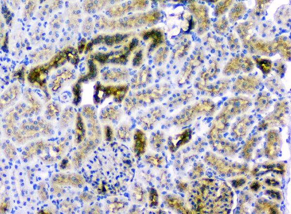
| WB, IHC, IF, ICC, FC |
|---|---|
| Primary Accession | Q9Y5K6 |
| Host | Mouse |
| Isotype | Mouse IgG1 |
| Reactivity | Rat, Human, Mouse |
| Clonality | Monoclonal |
| Format | Lyophilized |
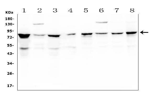
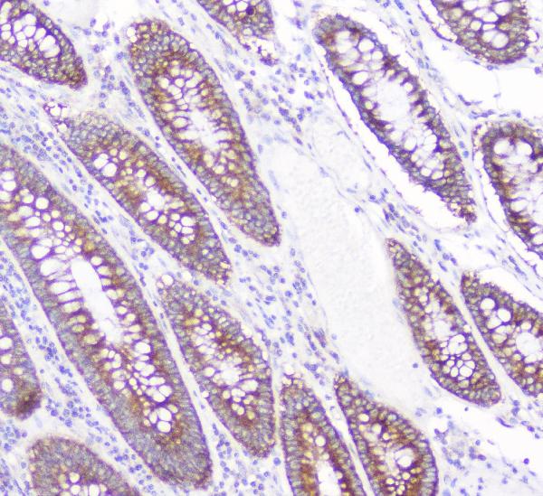
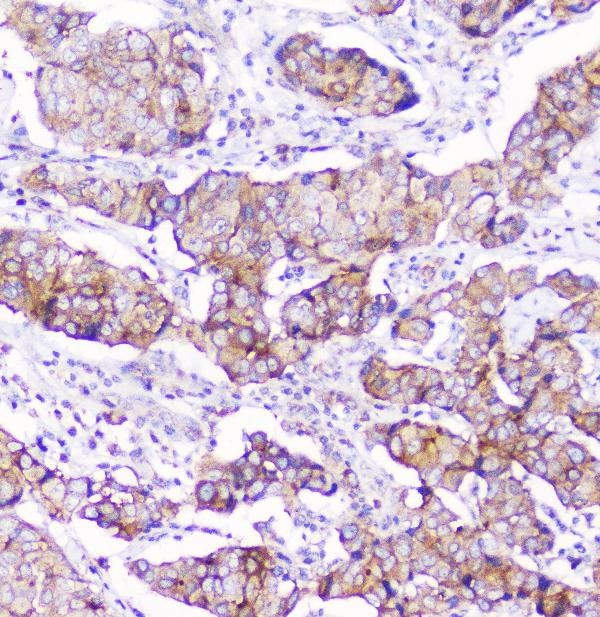
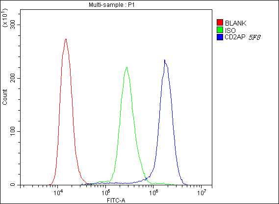
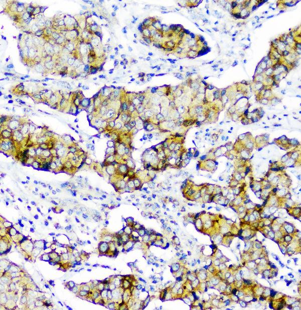
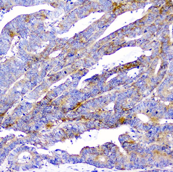
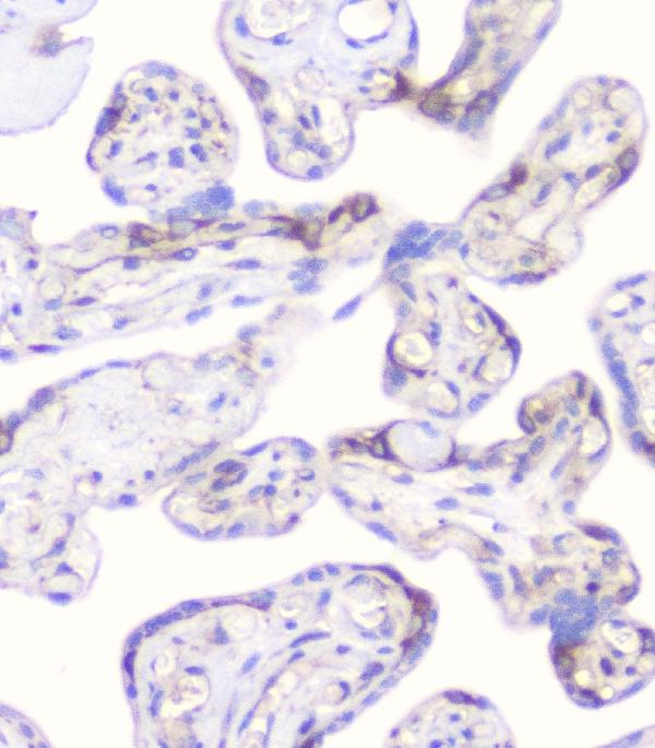
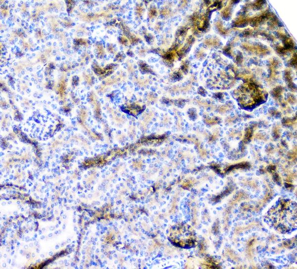
| Description | Anti-CD2AP Antibody Picoband™ (monoclonal, 5F8) . Tested in Flow Cytometry, IF, IHC, ICC, WB applications. This antibody reacts with Human, Mouse, Rat. |
| Gene ID | 23607 |
|---|---|
| Other Names | CD2-associated protein, Adapter protein CMS, Cas ligand with multiple SH3 domains, CD2AP |
| Calculated MW | 80 kDa |
| Application Details | Western blot, 0.1-0.5 µg/ml Immunohistochemistry (Paraffin-embedded Section), 0.5-1 µg/ml Immunocytochemistry/Immunofluorescence, 5 µg/ml Flow Cytometry, 1-3 µg/1x10^6 cells |
| Subcellular Localization | Cytoplasm, cytoskeleton. Cell projection, ruffle. Colocalizes with F-actin and BCAR1/p130Cas in membrane ruffles. Located at podocyte slit diaphragm between podocyte foot processes (By similarity). During late anaphase and telophase, concentrates in the vicinity of the midzone microtubules and in the midbody in late telophase. |
| Tissue Specificity | Widely expressed in fetal and adult tissues. |
| Contents | Each vial contains 4mg Trehalose, 0.9mg NaCl, 0.2mg Na2HPO4, 0.05mg NaN3. |
| Clone Names | Clone: 5F8 |
| Immunogen | E. coli-derived human CD2AP recombinant protein (Position: K253-K337). |
| Cross Reactivity | No cross-reactivity with other proteins. |
| Storage | Store at -20˚C for one year from date of receipt. After reconstitution, at 4˚C for one month. It can also be aliquotted and stored frozen at -20˚C for six months. Avoid repeated freeze-thaw cycles. |
| Name | CD2AP |
|---|---|
| Function | Seems to act as an adapter protein between membrane proteins and the actin cytoskeleton (PubMed:10339567). In collaboration with CBLC, modulates the rate of RET turnover and may act as regulatory checkpoint that limits the potency of GDNF on neuronal survival. Controls CBLC function, converting it from an inhibitor to a promoter of RET degradation (By similarity). May play a role in receptor clustering and cytoskeletal polarity in the junction between T-cell and antigen-presenting cell (By similarity). May anchor the podocyte slit diaphragm to the actin cytoskeleton in renal glomerolus. Also required for cytokinesis (PubMed:15800069). Plays a role in epithelial cell junctions formation (PubMed:22891260). |
| Cellular Location | Cytoplasm, cytoskeleton. Cell projection, ruffle. Cell junction. Note=Colocalizes with F-actin and BCAR1/p130Cas in membrane ruffles (PubMed:10339567). Located at podocyte slit diaphragm between podocyte foot processes (By similarity). During late anaphase and telophase, concentrates in the vicinity of the midzone microtubules and in the midbody in late telophase (PubMed:15800069). {ECO:0000250|UniProtKB:Q9JLQ0, ECO:0000269|PubMed:10339567, ECO:0000269|PubMed:15800069} |
| Tissue Location | Widely expressed in fetal and adult tissues. |

Thousands of laboratories across the world have published research that depended on the performance of antibodies from Abcepta to advance their research. Check out links to articles that cite our products in major peer-reviewed journals, organized by research category.
info@abcepta.com, and receive a free "I Love Antibodies" mug.

Provided below are standard protocols that you may find useful for product applications.
Background
CD2-associated protein is a protein that in humans is encoded by the CD2AP gene. This gene encodes a scaffolding molecule that regulates the actin cytoskeleton. The protein directly interacts with filamentous actin and a variety of cell membrane proteins through multiple actin binding sites, SH3 domains, and a proline-rich region containing binding sites for SH3 domains. The cytoplasmic protein localizes to membrane ruffles, lipid rafts, and the leading edges of cells. It is implicated in dynamic actin remodeling and membrane trafficking that occurs during receptor endocytosis and cytokinesis. Haploinsufficiency of this gene is implicated in susceptibility to glomerular disease.
If you have used an Abcepta product and would like to share how it has performed, please click on the "Submit Review" button and provide the requested information. Our staff will examine and post your review and contact you if needed.
If you have any additional inquiries please email technical services at tech@abcepta.com.
Ordering Information
Other Products
Shipping Information
