


 Foundational characteristics of cancer include proliferation, angiogenesis, migration, evasion of apoptosis, and cellular immortality. Find key markers for these cellular processes and antibodies to detect them.
 The SUMOplot™ Analysis Program predicts and scores sumoylation sites in your protein. SUMOylation is a post-translational modification involved in various cellular processes, such as nuclear-cytosolic transport, transcriptional regulation, apoptosis, protein stability, response to stress, and progression through the cell cycle.
 The Autophagy Receptor Motif Plotter predicts and scores autophagy receptor binding sites in your protein. Identifying proteins connected to this pathway is critical to understanding the role of autophagy in physiological as well as pathological processes such as development, differentiation, neurodegenerative diseases, stress, infection, and cancer.
Anti-CASR Antibody Picoband™ (monoclonal, 11E9)
- SPECIFICATION
- CITATIONS
- PROTOCOLS
- BACKGROUND

Application
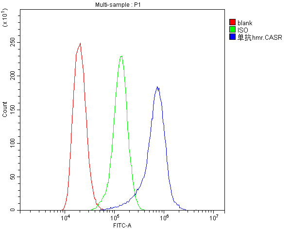
| WB, IHC, ICC, FC |
|---|---|
| Primary Accession | P41180 |
| Host | Mouse |
| Isotype | Mouse IgG2b |
| Reactivity | Human |
| Clonality | Monoclonal |
| Format | Lyophilized |
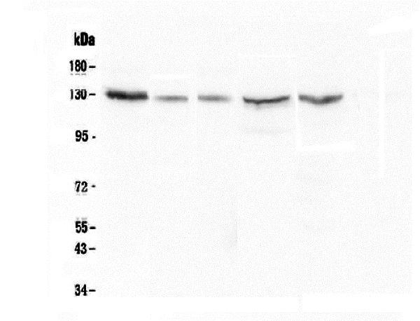
| Description | Anti-CASR Antibody Picoband™ (monoclonal, 11E9) . Tested in Flow Cytometry, IHC, ICC, WB applications. This antibody reacts with Human. |
| Reconstitution | Add 0.2ml of distilled water will yield a concentration of 500ug/ml. |
| Gene ID | 846 |
|---|---|
| Other Names | Extracellular calcium-sensing receptor, CaR, CaSR, hCasR, Parathyroid cell calcium-sensing receptor 1, PCaR1, CASR (HGNC:1514) |
| Calculated MW | 130 kDa |
| Application Details | Western blot, 0.1-0.5 µg/ml Immunohistochemistry (Paraffin-embedded Section), 0.5-1 µg/ml Immunohistochemistry (Frozen Section), 0.5-1 µg/ml Immunocytochemistry, 0.5-1 µg/ml Flow Cytometry, 1-3 µg/1x106 cells |
| Subcellular Localization | Cell membrane |
| Tissue Specificity | Expressed in the temporal lobe, frontal lobe, parietal lobe, hippocampus, and cerebellum. Also found in kidney, lung, liver, heart, skeletal muscle, placenta. |
| Contents | Each vial contains 4mg Trehalose, 0.9mg NaCl, 0.2mg Na2HPO4, 0.05mg NaN3. |
| Clone Names | Clone: 11E9 |
| Immunogen | E. coli-derived human CASR recombinant protein (Position: Q926-S1078). Human CASR shares 80.5% and 78.6% amino acid (aa) sequence identity with mouse and rat CASR, respectively. |
| Cross Reactivity | No cross-reactivity with other proteins. |
| Storage | Store at -20˚C for one year from date of receipt. After reconstitution, at 4˚C for one month. It can also be aliquotted and stored frozen at -20˚C for six months. Avoid repeated freeze-thaw cycles. |
| Name | CASR (HGNC:1514) |
|---|---|
| Function | G-protein-coupled receptor that senses changes in the extracellular concentration of calcium ions and plays a key role in maintaining calcium homeostasis (PubMed:17555508, PubMed:19789209, PubMed:21566075, PubMed:22114145, PubMed:22789683, PubMed:23966241, PubMed:25104082, PubMed:25292184, PubMed:25766501, PubMed:26386835, PubMed:7759551, PubMed:8636323, PubMed:8702647, PubMed:8878438). Senses fluctuations in the circulating calcium concentration and modulates the production of parathyroid hormone (PTH) in parathyroid glands (By similarity). The activity of this receptor is mediated by a G-protein that activates a phosphatidylinositol-calcium second messenger system (PubMed:7759551). The G-protein-coupled receptor activity is activated by a co-agonist mechanism: aromatic amino acids, such as Trp or Phe, act concertedly with divalent cations, such as calcium or magnesium, to achieve full receptor activation (PubMed:27386547, PubMed:27434672). |
| Cellular Location | Cell membrane; Multi-pass membrane protein |
| Tissue Location | Expressed in the temporal lobe, frontal lobe, parietal lobe, hippocampus, and cerebellum. Also found in kidney, lung, liver, heart, skeletal muscle, placenta. |

Thousands of laboratories across the world have published research that depended on the performance of antibodies from Abcepta to advance their research. Check out links to articles that cite our products in major peer-reviewed journals, organized by research category.
info@abcepta.com, and receive a free "I Love Antibodies" mug.

Provided below are standard protocols that you may find useful for product applications.
Background
The calcium-sensing receptor (CaSR) is a G protein-coupled receptor that is expressed in the parathyroid hormone (PTH)-producing chief cells of the parathyroid gland, and the cells lining the kidney tubule. It senses small changes in circulating calcium concentration and couples this information to intracellular signaling pathways that modify PTH secretion or renal cation handling, thus this protein plays an essential role in maintaining mineral ion homeostasis. Mutations in this gene cause familial hypocalciuric hypercalcemia, familial, isolated hypoparathyroidism, and neonatal severe primary hyperparathyroidism.
If you have used an Abcepta product and would like to share how it has performed, please click on the "Submit Review" button and provide the requested information. Our staff will examine and post your review and contact you if needed.
If you have any additional inquiries please email technical services at tech@abcepta.com.
Ordering Information
Other Products
Shipping Information
