


 Foundational characteristics of cancer include proliferation, angiogenesis, migration, evasion of apoptosis, and cellular immortality. Find key markers for these cellular processes and antibodies to detect them.
 The SUMOplot™ Analysis Program predicts and scores sumoylation sites in your protein. SUMOylation is a post-translational modification involved in various cellular processes, such as nuclear-cytosolic transport, transcriptional regulation, apoptosis, protein stability, response to stress, and progression through the cell cycle.
 The Autophagy Receptor Motif Plotter predicts and scores autophagy receptor binding sites in your protein. Identifying proteins connected to this pathway is critical to understanding the role of autophagy in physiological as well as pathological processes such as development, differentiation, neurodegenerative diseases, stress, infection, and cancer.
Anti-GSTM1 Antibody Picoband™ (monoclonal, 11F2)
- SPECIFICATION
- CITATIONS
- PROTOCOLS
- BACKGROUND

Application
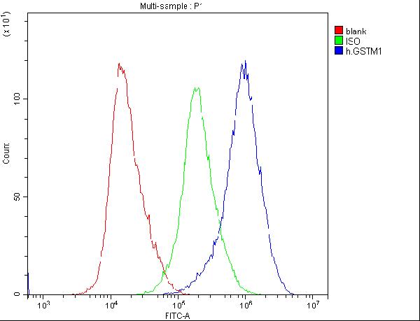
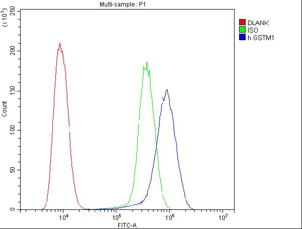
| WB, IHC, ICC, FC |
|---|---|
| Primary Accession | P09488 |
| Host | Mouse |
| Isotype | Mouse IgG1 |
| Reactivity | Rat, Human, Mouse |
| Clonality | Monoclonal |
| Format | Lyophilized |
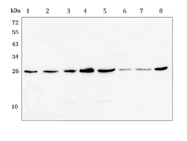
| Description | Anti-GSTM1 Antibody Picoband™ (monoclonal, 11F2) . Tested in Flow Cytometry, IHC, ICC, WB applications. This antibody reacts with Human, Mouse, Rat. |
| Reconstitution | Add 0.2ml of distilled water will yield a concentration of 500ug/ml. |
| Gene ID | 2944 |
|---|---|
| Other Names | Glutathione S-transferase Mu 1, 2.5.1.18, GST HB subunit 4, GST class-mu 1, GSTM1-1, GSTM1a-1a, GSTM1b-1b, GTH4, GSTM1 (HGNC:4632), GST1 |
| Calculated MW | 26 kDa |
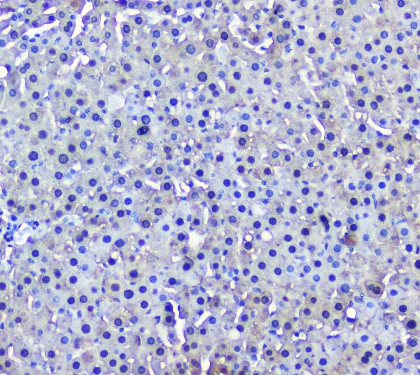
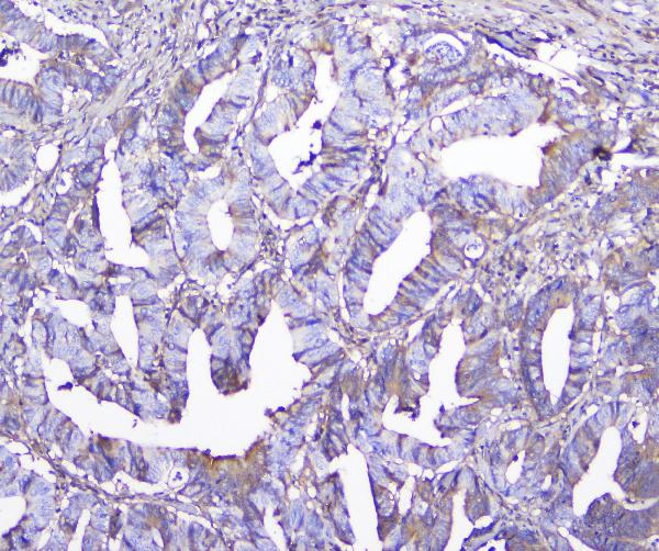
| Application Details | Western blot, 0.1-0.5 µg/ml Immunohistochemistry (Paraffin-embedded Section), 0.5-1 µg/ml Immunohistochemistry (Frozen Section), 0.5-1 µg/ml Immunocytochemistry, 0.5-1 µg/ml Flow Cytometry, 1-3 µg/1x106 cells |
| Subcellular Localization | Cytoplasm. |
| Tissue Specificity | Liver (at protein level). |
| Contents | Each vial contains 4mg Trehalose, 0.9mg NaCl, 0.2mg Na2HPO4, 0.05mg NaN3. |
| Clone Names | Clone: 11F2 |
| Immunogen | A synthetic peptide corresponding to a sequence in the middle region of human GSTM1, which shares 70.6% and 73.5% amino acid (aa) sequence identity with mouse and rat GSTM1, respectively. |
| Cross Reactivity | No cross-reactivity with other proteins. |
| Storage | Store at -20˚C for one year from date of receipt. After reconstitution, at 4˚C for one month. It can also be aliquotted and stored frozen at -20˚C for six months. Avoid repeated freeze-thaw cycles. |
| Name | GSTM1 (HGNC:4632) |
|---|---|
| Synonyms | GST1 |
| Function | Conjugation of reduced glutathione to a wide number of exogenous and endogenous hydrophobic electrophiles. Involved in the formation of glutathione conjugates of both prostaglandin A2 (PGA2) and prostaglandin J2 (PGJ2) (PubMed:9084911). Participates in the formation of novel hepoxilin regioisomers (PubMed:21046276). |
| Cellular Location | Cytoplasm. |
| Tissue Location | Liver (at protein level). |

Thousands of laboratories across the world have published research that depended on the performance of antibodies from Abcepta to advance their research. Check out links to articles that cite our products in major peer-reviewed journals, organized by research category.
info@abcepta.com, and receive a free "I Love Antibodies" mug.

Provided below are standard protocols that you may find useful for product applications.
Background
Glutathione S-transferase Mu 1 (gene name GSTM1) is a human glutathione S-transferase. Cytosolic and membrane-bound forms of glutathione S-transferase are encoded by two distinct supergene families. At present, eight distinct classes of the soluble cytoplasmic mammalian glutathione S-transferases have been identified: alpha, kappa, mu, omega, pi, sigma, theta and zeta. This gene encodes a glutathione S-transferase that belongs to the mu class. The mu class of enzymes functions in the detoxification of electrophilic compounds, including carcinogens, therapeutic drugs, environmental toxins and products of oxidative stress, by conjugation with glutathione. The genes encoding the mu class of enzymes are organized in a gene cluster on chromosome 1p13.3 and are known to be highly polymorphic. These genetic variations can change an individual's susceptibility to carcinogens and toxins as well as affect the toxicity and efficacy of certain drugs. Null mutations of this class mu gene have been linked with an increase in a number of cancers, likely due to an increased susceptibility to environmental toxins and carcinogens. Multiple protein isoforms are encoded by transcript variants of this gene.
If you have used an Abcepta product and would like to share how it has performed, please click on the "Submit Review" button and provide the requested information. Our staff will examine and post your review and contact you if needed.
If you have any additional inquiries please email technical services at tech@abcepta.com.
Ordering Information
Other Products
Shipping Information
