


 Foundational characteristics of cancer include proliferation, angiogenesis, migration, evasion of apoptosis, and cellular immortality. Find key markers for these cellular processes and antibodies to detect them.
 The SUMOplot™ Analysis Program predicts and scores sumoylation sites in your protein. SUMOylation is a post-translational modification involved in various cellular processes, such as nuclear-cytosolic transport, transcriptional regulation, apoptosis, protein stability, response to stress, and progression through the cell cycle.
 The Autophagy Receptor Motif Plotter predicts and scores autophagy receptor binding sites in your protein. Identifying proteins connected to this pathway is critical to understanding the role of autophagy in physiological as well as pathological processes such as development, differentiation, neurodegenerative diseases, stress, infection, and cancer.
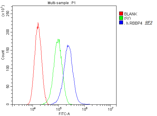
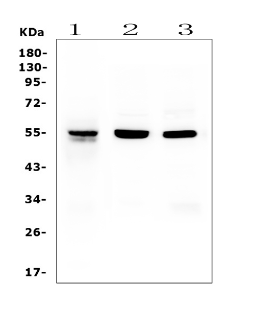
Anti-RbAp48 RBBP4 Antibody Picoband™ (monoclonal, 9F3)
- SPECIFICATION
- CITATIONS
- PROTOCOLS
- BACKGROUND

Application
| WB, IHC |
|---|---|
| Primary Accession | Q09028 |
| Host | Mouse |
| Isotype | Mouse IgG1 |
| Reactivity | Rat, Human, Mouse |
| Clonality | Monoclonal |
| Format | Lyophilized |
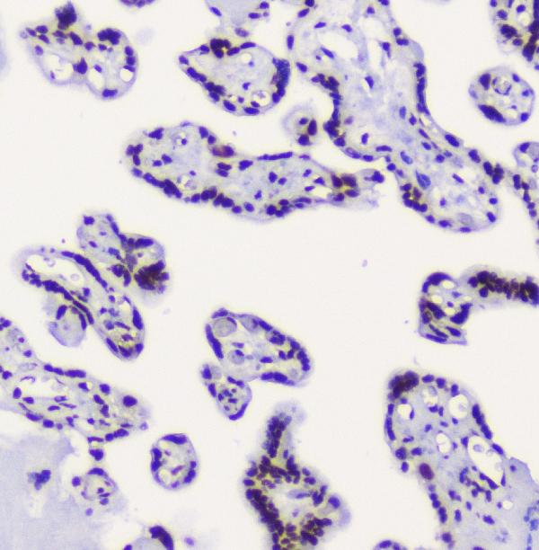
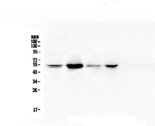
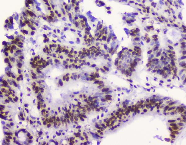
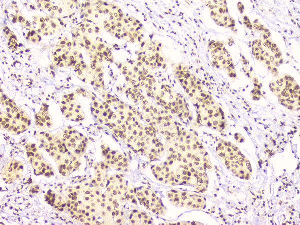
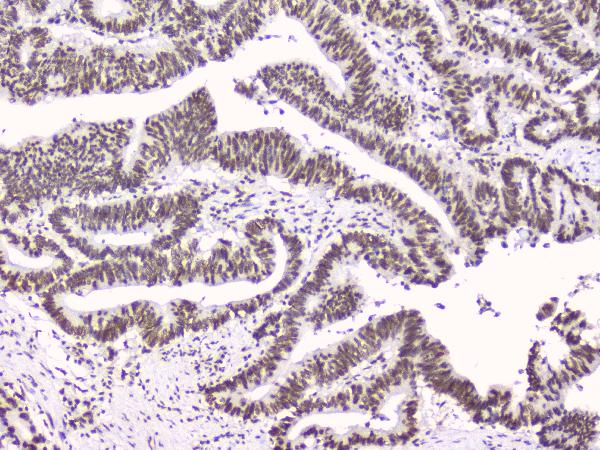
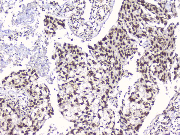
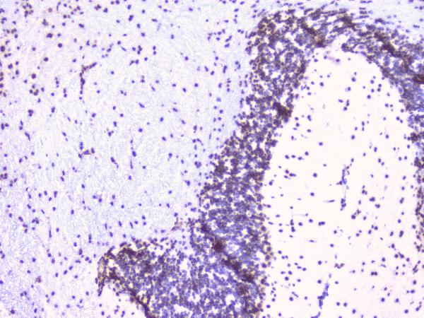
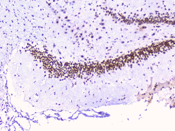
| Description | Anti-RbAp48 RBBP4 Antibody Picoband™ (monoclonal, 9F3) . Tested in IHC, WB applications. This antibody reacts with Human, Mouse, Rat. |
| Reconstitution | Add 0.2ml of distilled water will yield a concentration of 500ug/ml. |
| Gene ID | 5928 |
|---|---|
| Other Names | Histone-binding protein RBBP4, Chromatin assembly factor 1 subunit C, CAF-1 subunit C, Chromatin assembly factor I p48 subunit, CAF-I 48 kDa subunit, CAF-I p48, Nucleosome-remodeling factor subunit RBAP48, Retinoblastoma-binding protein 4, RBBP-4, Retinoblastoma-binding protein p48, RBBP4, RBAP48 |
| Calculated MW | 55 kDa |
| Application Details | Western blot, 0.1-0.5 µg/ml Immunohistochemistry (Paraffin-embedded Section), 0.5-1 µg/ml |
| Subcellular Localization | Nucleus. |
| Contents | Each vial contains 4mg Trehalose, 0.9mg NaCl, 0.2mg Na2HPO4, 0.05mg NaN3. |
| Clone Names | Clone: 9F3 |
| Immunogen | A synthetic peptide corresponding to a sequence at the C-terminus of human RbAp48, identical to the related mouse sequence. |
| Cross Reactivity | No cross-reactivity with other proteins. |
| Storage | Store at -20˚C for one year from date of receipt. After reconstitution, at 4˚C for one month. It can also be aliquotted and stored frozen at -20˚C for six months. Avoid repeated freeze-thaw cycles. |
| Name | RBBP4 |
|---|---|
| Synonyms | RBAP48 |
| Function | Core histone-binding subunit that may target chromatin assembly factors, chromatin remodeling factors and histone deacetylases to their histone substrates in a manner that is regulated by nucleosomal DNA (PubMed:10866654). Component of the chromatin assembly factor 1 (CAF-1) complex, which is required for chromatin assembly following DNA replication and DNA repair (PubMed:8858152). Component of the core histone deacetylase (HDAC) complex, which promotes histone deacetylation and consequent transcriptional repression (PubMed:9150135). Component of the nucleosome remodeling and histone deacetylase complex (the NuRD complex), which promotes transcriptional repression by histone deacetylation and nucleosome remodeling (PubMed:16428440, PubMed:28977666, PubMed:39460621). Component of the PRC2 complex, which promotes repression of homeotic genes during development (PubMed:29499137, PubMed:31959557). Component of the NURF (nucleosome remodeling factor) complex (PubMed:14609955, PubMed:15310751). |
| Cellular Location | Nucleus. Chromosome, telomere. Note=Localizes to chromatin as part of the PRC2 complex. |
| Tissue Location | Expressed in neuroblastoma cells. |

Thousands of laboratories across the world have published research that depended on the performance of antibodies from Abcepta to advance their research. Check out links to articles that cite our products in major peer-reviewed journals, organized by research category.
info@abcepta.com, and receive a free "I Love Antibodies" mug.

Provided below are standard protocols that you may find useful for product applications.
Background
Histone-binding protein RBBP4 (also known as RbAp48, or NURF55) is a protein that in humans is encoded by the RBBP4 gene. This gene encodes a ubiquitously expressed nuclear protein which belongs to a highly conserved subfamily of WD-repeat proteins. It is present in protein complexes involved in histone acetylation and chromatin assembly. And it is part of the Mi-2 complex which has been implicated in chromatin remodeling and transcriptional repression associated with histone deacetylation. This encoded protein is also part of co-repressor complexes, which is an integral component of transcriptional silencing. It is found among several cellular proteins that bind directly to retinoblastoma protein to regulate cell proliferation. This protein also seems to be involved in transcriptional repression of E2F-responsive genes. Three transcript variants encoding different isoforms have been found for this gene.
If you have used an Abcepta product and would like to share how it has performed, please click on the "Submit Review" button and provide the requested information. Our staff will examine and post your review and contact you if needed.
If you have any additional inquiries please email technical services at tech@abcepta.com.
Ordering Information
Other Products
Shipping Information
