


 Foundational characteristics of cancer include proliferation, angiogenesis, migration, evasion of apoptosis, and cellular immortality. Find key markers for these cellular processes and antibodies to detect them.
 The SUMOplot™ Analysis Program predicts and scores sumoylation sites in your protein. SUMOylation is a post-translational modification involved in various cellular processes, such as nuclear-cytosolic transport, transcriptional regulation, apoptosis, protein stability, response to stress, and progression through the cell cycle.
 The Autophagy Receptor Motif Plotter predicts and scores autophagy receptor binding sites in your protein. Identifying proteins connected to this pathway is critical to understanding the role of autophagy in physiological as well as pathological processes such as development, differentiation, neurodegenerative diseases, stress, infection, and cancer.
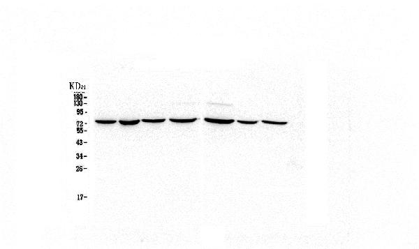
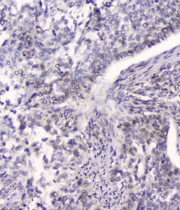
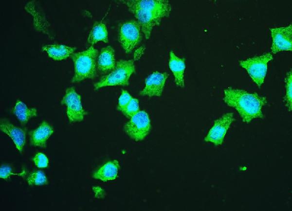
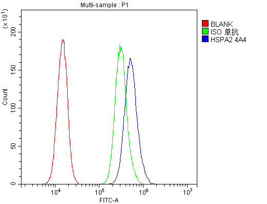
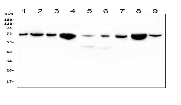
Anti-HSPA2 Antibody Picoband™ (monoclonal, 4A4)
- SPECIFICATION
- CITATIONS
- PROTOCOLS
- BACKGROUND

Application
| WB, IHC, IF, ICC, FC |
|---|---|
| Primary Accession | P54652 |
| Host | Mouse |
| Isotype | Mouse IgG1 |
| Reactivity | Rat, Human, Mouse |
| Clonality | Monoclonal |
| Format | Lyophilized |
| Description | Anti-HSPA2 Antibody Picoband™ (monoclonal, 4A4) . Tested in Flow Cytometry, IF, IHC, ICC, WB applications. This antibody reacts with Human, Mouse, Rat. |
| Reconstitution | Add 0.2ml of distilled water will yield a concentration of 500ug/ml. |
| Gene ID | 3306 |
|---|---|
| Other Names | Heat shock-related 70 kDa protein 2, Heat shock 70 kDa protein 2, Heat shock protein family A member 2, HSPA2 |
| Calculated MW | 70 kDa |
| Application Details | Western blot, 0.1-0.5 µg/ml Immunohistochemistry (Paraffin-embedded Section), 0.5-1 µg/ml Immunocytochemistry/Immunofluorescence, 2 µg/ml Flow Cytometry, 1-3 µg/1x10^6 cells |
| Subcellular Localization | Cytoplasm, cytoskeleton, spindle |
| Contents | Each vial contains 4mg Trehalose, 0.9mg NaCl, 0.2mg Na2HPO4, 0.05mg NaN3. |
| Clone Names | Clone: 4A4 |
| Immunogen | A synthetic peptide corresponding to a sequence at the C-terminus of human HSPA2, identical to the related mouse and rat sequences. |
| Cross Reactivity | No cross-reactivity with other proteins. |
| Storage | Store at -20˚C for one year from date of receipt. After reconstitution, at 4˚C for one month. It can also be aliquotted and stored frozen at -20˚C for six months. Avoid repeated freeze-thaw cycles. |
| Name | HSPA2 |
|---|---|
| Function | Molecular chaperone implicated in a wide variety of cellular processes, including protection of the proteome from stress, folding and transport of newly synthesized polypeptides, activation of proteolysis of misfolded proteins and the formation and dissociation of protein complexes. Plays a pivotal role in the protein quality control system, ensuring the correct folding of proteins, the re-folding of misfolded proteins and controlling the targeting of proteins for subsequent degradation. This is achieved through cycles of ATP binding, ATP hydrolysis and ADP release, mediated by co-chaperones. The affinity for polypeptides is regulated by its nucleotide bound state. In the ATP-bound form, it has a low affinity for substrate proteins. However, upon hydrolysis of the ATP to ADP, it undergoes a conformational change that increases its affinity for substrate proteins. It goes through repeated cycles of ATP hydrolysis and nucleotide exchange, which permits cycles of substrate binding and release (PubMed:26865365). Plays a role in spermatogenesis. In association with SHCBP1L may participate in the maintenance of spindle integrity during meiosis in male germ cells (By similarity). |
| Cellular Location | Cytoplasm, cytoskeleton, spindle {ECO:0000250|UniProtKB:P17156}. Note=Colocalizes with SHCBP1L at spindle during the meiosis process. {ECO:0000250|UniProtKB:P17156} |

Thousands of laboratories across the world have published research that depended on the performance of antibodies from Abcepta to advance their research. Check out links to articles that cite our products in major peer-reviewed journals, organized by research category.
info@abcepta.com, and receive a free "I Love Antibodies" mug.

Provided below are standard protocols that you may find useful for product applications.
Background
HSPA2 (heat shock 70kDa protein 2) is also known as HEAT-SHOCK PROTEIN, 70-KD, 2, HSP70-2, HEAT-SHOCK PROTEIN, 70-KD, 3 or HSP70-3. Analysis of the sequence indicated that HSPA2 is the human homolog of the murine Hsp70-2 gene, with 91.7% identity in the nucleotide coding sequence and 98.2% in the corresponding amino acid sequence. HSPA2 has less amino acid homology to the other members of the human HSP70 gene family. HSPA2 is constitutively expressed in most tissues, with very high levels in testis and skeletal muscle. The HSPA2 gene is located on chromosome 14q22-q24. Immunohistochemical analysis detected weak expression of HSPA2 in spermatocytes and stronger expression in spermatids and in the tail of mature sperm. HSPA2 may be critical to sperm maturation through its role as a protein chaperone.
If you have used an Abcepta product and would like to share how it has performed, please click on the "Submit Review" button and provide the requested information. Our staff will examine and post your review and contact you if needed.
If you have any additional inquiries please email technical services at tech@abcepta.com.
Ordering Information
Other Products
Shipping Information
