


 Foundational characteristics of cancer include proliferation, angiogenesis, migration, evasion of apoptosis, and cellular immortality. Find key markers for these cellular processes and antibodies to detect them.
 The SUMOplot™ Analysis Program predicts and scores sumoylation sites in your protein. SUMOylation is a post-translational modification involved in various cellular processes, such as nuclear-cytosolic transport, transcriptional regulation, apoptosis, protein stability, response to stress, and progression through the cell cycle.
 The Autophagy Receptor Motif Plotter predicts and scores autophagy receptor binding sites in your protein. Identifying proteins connected to this pathway is critical to understanding the role of autophagy in physiological as well as pathological processes such as development, differentiation, neurodegenerative diseases, stress, infection, and cancer.
> home > Products > Primary Antibodies > Antibody Collections > Pancreas positive > Anti-TRIM25 Monoclonal Antibody
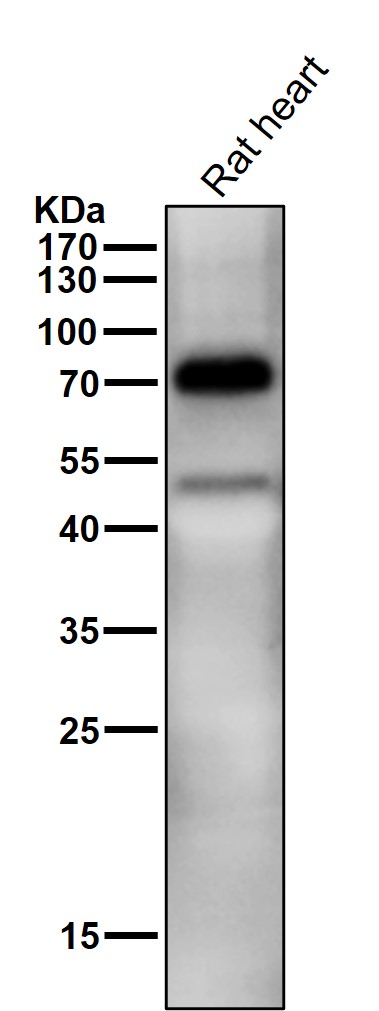
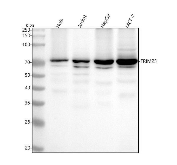
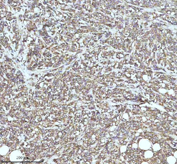
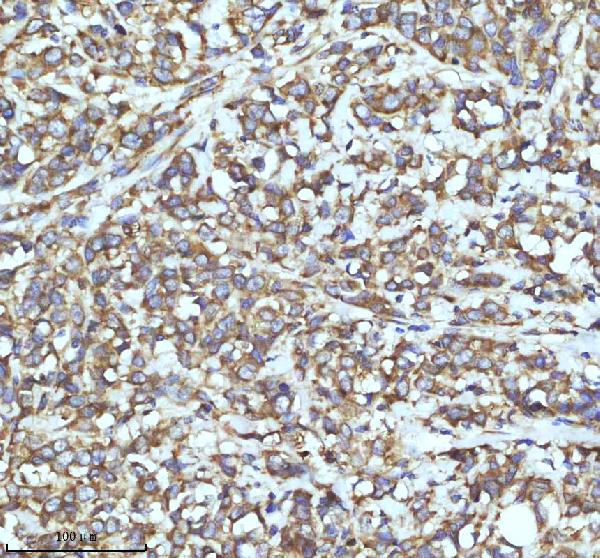
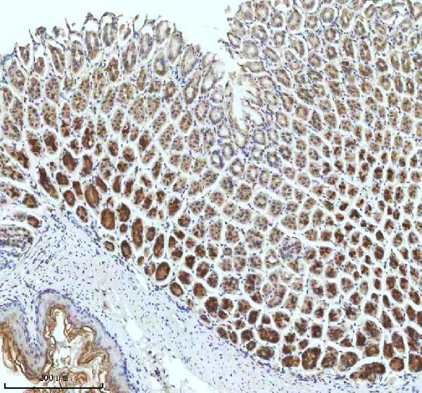
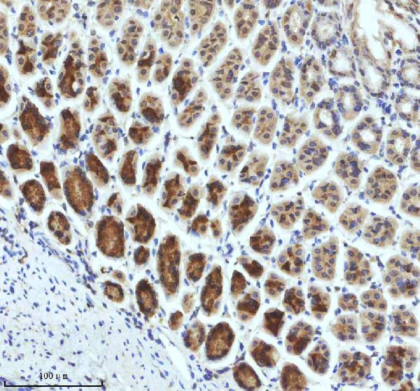
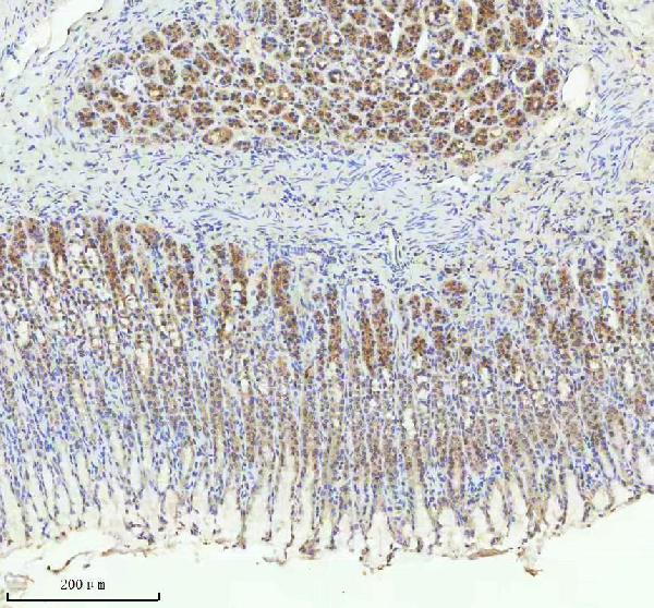
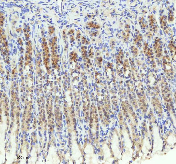
Anti-TRIM25 Monoclonal Antibody
- SPECIFICATION
- CITATIONS
- PROTOCOLS
- BACKGROUND

Application
| WB, IHC, IF, ICC, IP, FC |
|---|---|
| Primary Accession | Q14258 |
| Host | Rabbit |
| Isotype | Rabbit IgG |
| Reactivity | Rat, Human, Mouse |
| Clonality | Monoclonal |
| Format | Liquid |
| Description | Anti-TRIM25 Monoclonal Antibody . Tested in WB, IHC, ICC/IF, IP, Flow Cytometry applications. This antibody reacts with Human, Mouse, Rat. |
| Gene ID | 7706 |
|---|---|
| Other Names | E3 ubiquitin/ISG15 ligase TRIM25, 6.3.2.n3, Estrogen-responsive finger protein, RING finger protein 147, RING-type E3 ubiquitin transferase, 2.3.2.27, RING-type E3 ubiquitin transferase TRIM25, Tripartite motif-containing protein 25, Ubiquitin/ISG15-conjugating enzyme TRIM25, Zinc finger protein 147, TRIM25, EFP {ECO:0000303|PubMed:8248217}, RNF147, ZNF147 |
| Calculated MW | 70973 Da |
| Application Details | WB 1:1000-1:5000 IHC 1:50-1:200 ICC/IF 1:100-1:500 IP 1:50 FC 1:60 |
| Contents | Rabbit IgG in phosphate buffered saline, pH 7.4, 150mM NaCl, 0.02% sodium azide and 50% glycerol, 0.4-0.5mg/ml BSA. |
| Clone Names | Clone: ADFG-20 |
| Immunogen | A synthesized peptide derived from human TRIM25 Functions as an ubiquitin E3 ligase and as an ISG15 E3 ligase. Involved in innate immune defense against viruses by mediating ubiquitination of DDX58. Mediates 'Lys-63'-linked polyubiquitination of the DDX58 N-terminal CARD-like region which is crucial for triggering the cytosolic signal transduction that leads to the production of interferons in response to viral infection. |
| Purification | Affinity-chromatography |
| Storage | Store at -20°C for one year. For short term storage and frequent use, store at 4°C for up to one month. Avoid repeated freeze-thaw cycles. |
| Name | TRIM25 |
|---|---|
| Synonyms | EFP {ECO:0000303|PubMed:8248217}, RNF147 |
| Function | Functions as a ubiquitin E3 ligase and as an ISG15 E3 ligase (PubMed:16352599). Involved in innate immune defense against viruses by mediating ubiquitination of RIGI and IFIH1 (PubMed:17392790, PubMed:29357390, PubMed:30193849, PubMed:31710640, PubMed:33849980, PubMed:36045682). Mediates 'Lys-63'-linked polyubiquitination of the RIGI N-terminal CARD-like region and may play a role in signal transduction that leads to the production of interferons in response to viral infection (PubMed:17392790, PubMed:23950712). Mediates 'Lys-63'- linked polyubiquitination of IFIH1 (PubMed:30193849). Promotes ISGylation of 14-3-3 sigma (SFN), an adapter protein implicated in the regulation of a large spectrum signaling pathway (PubMed:16352599, PubMed:17069755). Mediates estrogen action in various target organs (PubMed:22452784). Mediates the ubiquitination and subsequent proteasomal degradation of ZFHX3 (PubMed:22452784). Plays a role in promoting the restart of stalled replication forks via interaction with the KHDC3L-OOEP scaffold and subsequent ubiquitination of BLM, resulting in the recruitment and retainment of BLM at DNA replication forks (By similarity). Plays an essential role in the antiviral activity of ZAP/ZC3HAV1; an antiviral protein which inhibits the replication of certain viruses. Mechanistically, mediates 'Lys-63'- linked polyubiquitination of ZAP/ZC3HAV1 that is required for its optimal binding to target mRNA (PubMed:28060952, PubMed:28202764). Mediates also the ubiquitination of various substrates implicated in stress granule formation, nonsense-mediated mRNA decay, nucleoside synthesis and mRNA translation and stability (PubMed:36067236). |
| Cellular Location | Cytoplasm. Cytoplasm, Stress granule. Nucleus {ECO:0000250|UniProtKB:Q61510} |
| Tissue Location | Expressed in breast tumors (at protein level). Ubiquitous. |
Research Areas
Citations (0)

Thousands of laboratories across the world have published research that depended on the performance of antibodies from Abcepta to advance their research. Check out links to articles that cite our products in major peer-reviewed journals, organized by research category.
Submit your citation using an Abcepta antibody to
info@abcepta.com, and receive a free "I Love Antibodies" mug.
info@abcepta.com, and receive a free "I Love Antibodies" mug.

Application Protocols
Provided below are standard protocols that you may find useful for product applications.
Abcepta welcomes feedback from its customers.
If you have used an Abcepta product and would like to share how it has performed, please click on the "Submit Review" button and provide the requested information. Our staff will examine and post your review and contact you if needed.
If you have any additional inquiries please email technical services at tech@abcepta.com.
$ 370.00
Cat# ABO14553
Ordering Information
United States
AlbaniaAustraliaAustriaBelgiumBosnia & HerzegovinaBrazilBulgariaCanadaCentral AmericaChinaCroatiaCyprusCzech RepublicDenmarkEstoniaFinlandFranceGermanyGreeceHong KongHungaryIcelandIndiaIndonesiaIrelandIsraelItalyJapanLatviaLithuaniaLuxembourgMacedoniaMalaysiaMaltaNetherlandsNew ZealandNorwayPakistanPolandPortugalRomaniaSerbiaSingaporeSlovakiaSloveniaSouth AfricaSouth KoreaSpainSwedenSwitzerlandTaiwanTurkeyUnited KingdomUnited StatesVietnamWorldwideOthersMexico
USA Headquarters
(888) 735-7227 / (858) 622-0099 or (858) 875-1900
Other Products
Shipping Information
Domestic orders (in stock items)
Shipped out the same day. Orders placed after 1 PM (PST) will ship out the next business day.
International orders
Contact your local distributors
