


 Foundational characteristics of cancer include proliferation, angiogenesis, migration, evasion of apoptosis, and cellular immortality. Find key markers for these cellular processes and antibodies to detect them.
 The SUMOplot™ Analysis Program predicts and scores sumoylation sites in your protein. SUMOylation is a post-translational modification involved in various cellular processes, such as nuclear-cytosolic transport, transcriptional regulation, apoptosis, protein stability, response to stress, and progression through the cell cycle.
 The Autophagy Receptor Motif Plotter predicts and scores autophagy receptor binding sites in your protein. Identifying proteins connected to this pathway is critical to understanding the role of autophagy in physiological as well as pathological processes such as development, differentiation, neurodegenerative diseases, stress, infection, and cancer.
> home > Products > Primary Antibodies > Antibody Collections > Prostate positive > Anti-PAK2 Monoclonal Antibody
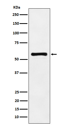
Anti-PAK2 Monoclonal Antibody
- SPECIFICATION
- CITATIONS
- PROTOCOLS
- BACKGROUND

Application
| WB, IHC, IF, ICC, FC |
|---|---|
| Primary Accession | Q13177 |
| Host | Rabbit |
| Isotype | Rabbit IgG |
| Reactivity | Rat, Human, Mouse |
| Clonality | Monoclonal |
| Format | Liquid |
| Description | Anti-PAK2 Monoclonal Antibody . Tested in WB, IHC, ICC/IF, Flow Cytometry applications. This antibody reacts with Human, Mouse, Rat. |
| Gene ID | 5062 |
|---|---|
| Other Names | Serine/threonine-protein kinase PAK 2, 2.7.11.1, Gamma-PAK, PAK65, S6/H4 kinase, p21-activated kinase 2, PAK-2, p58, PAK-2p27, p27, PAK-2p34, p34, C-t-PAK2, PAK2 |
| Calculated MW | 58043 Da |
| Application Details | WB 1:500-1:2000 IHC 1:100-1:500 ICC/IF 1:50-1:200 FC 1:80 |
| Contents | Rabbit IgG in phosphate buffered saline, pH 7.4, 150mM NaCl, 0.02% sodium azide and 50% glycerol, 0.4-0.5mg/ml BSA. |
| Clone Names | Clone: ADFE-16 |
| Immunogen | A synthesized peptide derived from human PAK2 The activated kinase acts on a variety of targets. Phosphorylates ribosomal protein S6, histone H4 and myelin basic protein. Full length PAK 2 stimulates cell survival and cell growth. The process is, at least in part, mediated by phosphorylation and inhibition of pro-apoptotic BAD. |
| Purification | Affinity-chromatography |
| Storage | Store at -20°C for one year. For short term storage and frequent use, store at 4°C for up to one month. Avoid repeated freeze-thaw cycles. |
| Name | PAK2 |
|---|---|
| Function | Serine/threonine protein kinase that plays a role in a variety of different signaling pathways including cytoskeleton regulation, cell motility, cell cycle progression, apoptosis or proliferation (PubMed:12853446, PubMed:16617111, PubMed:19273597, PubMed:19923322, PubMed:33693784, PubMed:7744004, PubMed:9171063). Acts as a downstream effector of the small GTPases CDC42 and RAC1 (PubMed:7744004). Activation by the binding of active CDC42 and RAC1 results in a conformational change and a subsequent autophosphorylation on several serine and/or threonine residues (PubMed:7744004). Full- length PAK2 stimulates cell survival and cell growth (PubMed:7744004). Phosphorylates MAPK4 and MAPK6 and activates the downstream target MAPKAPK5, a regulator of F-actin polymerization and cell migration (PubMed:21317288). Phosphorylates JUN and plays an important role in EGF-induced cell proliferation (PubMed:21177766). Phosphorylates many other substrates including histone H4 to promote assembly of H3.3 and H4 into nucleosomes, BAD, ribosomal protein S6, or MBP (PubMed:21724829). Phosphorylates CASP7, thereby preventing its activity (PubMed:21555521, PubMed:27889207). Additionally, associates with ARHGEF7 and GIT1 to perform kinase-independent functions such as spindle orientation control during mitosis (PubMed:19273597, PubMed:19923322). On the other hand, apoptotic stimuli such as DNA damage lead to caspase-mediated cleavage of PAK2, generating PAK-2p34, an active p34 fragment that translocates to the nucleus and promotes cellular apoptosis involving the JNK signaling pathway (PubMed:12853446, PubMed:16617111, PubMed:9171063). Caspase-activated PAK2 phosphorylates MKNK1 and reduces cellular translation (PubMed:15234964). |
| Cellular Location | [Serine/threonine-protein kinase PAK 2]: Cytoplasm Nucleus Note=MYO18A mediates the cellular distribution of the PAK2-ARHGEF7-GIT1 complex to the inner surface of the cell membrane |
| Tissue Location | Ubiquitously expressed. Higher levels seen in skeletal muscle, ovary, thymus and spleen |
Research Areas
Citations (0)

Thousands of laboratories across the world have published research that depended on the performance of antibodies from Abcepta to advance their research. Check out links to articles that cite our products in major peer-reviewed journals, organized by research category.
Submit your citation using an Abcepta antibody to
info@abcepta.com, and receive a free "I Love Antibodies" mug.
info@abcepta.com, and receive a free "I Love Antibodies" mug.

Application Protocols
Provided below are standard protocols that you may find useful for product applications.
Abcepta welcomes feedback from its customers.
If you have used an Abcepta product and would like to share how it has performed, please click on the "Submit Review" button and provide the requested information. Our staff will examine and post your review and contact you if needed.
If you have any additional inquiries please email technical services at tech@abcepta.com.
$ 370.00
Cat# ABO14551
Ordering Information
United States
AlbaniaAustraliaAustriaBelgiumBosnia & HerzegovinaBrazilBulgariaCanadaCentral AmericaChinaCroatiaCyprusCzech RepublicDenmarkEstoniaFinlandFranceGermanyGreeceHong KongHungaryIcelandIndiaIndonesiaIrelandIsraelItalyJapanLatviaLithuaniaLuxembourgMacedoniaMalaysiaMaltaNetherlandsNew ZealandNorwayPakistanPolandPortugalRomaniaSerbiaSingaporeSlovakiaSloveniaSouth AfricaSouth KoreaSpainSwedenSwitzerlandTaiwanTurkeyUnited KingdomUnited StatesVietnamWorldwideOthersMexico
USA Headquarters
(888) 735-7227 / (858) 622-0099 or (858) 875-1900
Other Products
Shipping Information
Domestic orders (in stock items)
Shipped out the same day. Orders placed after 1 PM (PST) will ship out the next business day.
International orders
Contact your local distributors
