


 Foundational characteristics of cancer include proliferation, angiogenesis, migration, evasion of apoptosis, and cellular immortality. Find key markers for these cellular processes and antibodies to detect them.
 The SUMOplot™ Analysis Program predicts and scores sumoylation sites in your protein. SUMOylation is a post-translational modification involved in various cellular processes, such as nuclear-cytosolic transport, transcriptional regulation, apoptosis, protein stability, response to stress, and progression through the cell cycle.
 The Autophagy Receptor Motif Plotter predicts and scores autophagy receptor binding sites in your protein. Identifying proteins connected to this pathway is critical to understanding the role of autophagy in physiological as well as pathological processes such as development, differentiation, neurodegenerative diseases, stress, infection, and cancer.
Anti-Dynamin 2 Monoclonal Antibody
- SPECIFICATION
- CITATIONS
- PROTOCOLS
- BACKGROUND

Application
| WB, IHC, IF, ICC, FC |
|---|---|
| Primary Accession | P50570 |
| Host | Rabbit |
| Isotype | Rabbit IgG |
| Reactivity | Rat, Human, Mouse |
| Clonality | Monoclonal |
| Format | Liquid |
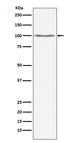
| Description | Anti-Dynamin 2 Monoclonal Antibody . Tested in WB, IHC, ICC/IF, Flow Cytometry applications. This antibody reacts with Human, Mouse, Rat. |
| Gene ID | 1785 |
|---|---|
| Other Names | Dynamin-2, 3.6.5.5, Dynamin 2, Dynamin II, DNM2 (HGNC:2974), DYN2 |
| Calculated MW | 98064 Da |
| Application Details | WB 1:500-1:1000 IHC 1:50-1:200 ICC/IF 1:50-1:200 FC 1:100 |
| Contents | Rabbit IgG in phosphate buffered saline, pH 7.4, 150mM NaCl, 0.02% sodium azide and 50% glycerol, 0.4-0.5mg/ml BSA. |
| Clone Names | Clone: ACFF-4 |
| Immunogen | A synthesized peptide derived from human Dynamin 2 Microtubule-associated force-producing protein involved in producing microtubule bundles and able to bind and hydrolyze GTP. Most probably involved in vesicular trafficking processes, in particular endocytosis. |
| Purification | Affinity-chromatography |
| Storage | Store at -20°C for one year. For short term storage and frequent use, store at 4°C for up to one month. Avoid repeated freeze-thaw cycles. |
| Name | DNM2 (HGNC:2974) |
|---|---|
| Synonyms | DYN2 |
| Function | Catalyzes the hydrolysis of GTP and utilizes this energy to mediate vesicle scission at plasma membrane during endocytosis and filament remodeling at many actin structures during organization of the actin cytoskeleton (PubMed:15731758, PubMed:19605363, PubMed:19623537, PubMed:33713620, PubMed:34744632). Plays an important role in vesicular trafficking processes, namely clathrin-mediated endocytosis (CME), exocytic and clathrin-coated vesicle from the trans-Golgi network, and PDGF stimulated macropinocytosis (PubMed:15731758, PubMed:19623537, PubMed:33713620). During vesicular trafficking process, associates to the membrane, through lipid binding, and self-assembles into ring-like structure through oligomerization to form a helical polymer around the vesicle membrane and leading to vesicle scission (PubMed:17636067, PubMed:34744632, PubMed:36445308). Plays a role in organization of the actin cytoskeleton by mediating arrangement of stress fibers and actin bundles in podocytes (By similarity). During organization of the actin cytoskeleton, self-assembles into ring-like structure that directly bundles actin filaments to form typical membrane tubules decorated with dynamin spiral polymers (By similarity). Self-assembly increases GTPase activity and the GTP hydrolysis causes the rapid depolymerization of dynamin spiral polymers, and results in dispersion of actin bundles (By similarity). Remodels, through its interaction with CTTN, bundled actin filaments in a GTPase-dependent manner and plays a role in orchestrating the global actomyosin cytoskeleton (PubMed:19605363). The interaction with CTTN stabilizes the interaction of DNM2 and actin filaments and stimulates the intrinsic GTPase activity that results in actin filament-barbed ends and increases the sensitivity of filaments in bundles to the actin depolymerizing factor, CFL1 (By similarity). Plays a role in the autophagy process, by participating in the formation of ATG9A vesicles destined for the autophagosomes through its interaction with SNX18 (PubMed:29437695), by mediating recycling endosome scission leading to autophagosome release through MAP1LC3B interaction (PubMed:29437695, PubMed:32315611). Also regulates maturation of apoptotic cell corpse-containing phagosomes by recruiting PIK3C3 to the phagosome membrane (By similarity). Also plays a role in cytokinesis (By similarity). May participate in centrosome cohesion through its interaction with TUBG1 (By similarity). Plays a role in the regulation of neuron morphology, axon growth and formation of neuronal growth cones (By similarity). Involved in membrane tubulation (PubMed:24135484). |
| Cellular Location | Cytoplasm, cytoskeleton. Cytoplasmic vesicle, clathrin-coated vesicle. Cell projection, uropodium. Endosome Cytoplasm, cytoskeleton, microtubule organizing center, centrosome. Cytoplasm, cytoskeleton, microtubule organizing center, centrosome, centriole Recycling endosome. Cell projection, phagocytic cup {ECO:0000250|UniProtKB:P39054}. Cytoplasmic vesicle, phagosome membrane {ECO:0000250|UniProtKB:P39054}; Peripheral membrane protein {ECO:0000250|UniProtKB:P39054}. Cell projection, podosome {ECO:0000250|UniProtKB:P39054}. Cytoplasm {ECO:0000250|UniProtKB:P39052}. Cell junction {ECO:0000250|UniProtKB:P39052}. Postsynaptic density {ECO:0000250|UniProtKB:P39052}. Synapse, synaptosome {ECO:0000250|UniProtKB:P39052}. Midbody {ECO:0000250|UniProtKB:P39052} Membrane, clathrin-coated pit {ECO:0000250|UniProtKB:P39052} Note=Localized in recycling endosomes fragment to release nascent autophagosomes (PubMed:32315611). Co-localizes with PIK3C3 and RAB5A to the nascent phagosome. Localized at focal ahesion site upon induction of focal adhesions and stress-fiber formation, when interacts with SDC4 (By similarity). Exists as a dynamic component of the centrosome Associates with clathrin-coated vesicles at both the plasma membrane and the trans-Golgi network (TGN) (By similarity) {ECO:0000250|UniProtKB:P39052, ECO:0000250|UniProtKB:P39054, ECO:0000269|PubMed:32315611} |
| Tissue Location | Widely expressed (PubMed:7590285). Expressed in skeletal muscle and the peripheral nerve (PubMed:19623537) |
Research Areas
Citations (0)

Thousands of laboratories across the world have published research that depended on the performance of antibodies from Abcepta to advance their research. Check out links to articles that cite our products in major peer-reviewed journals, organized by research category.
Submit your citation using an Abcepta antibody to
info@abcepta.com, and receive a free "I Love Antibodies" mug.
info@abcepta.com, and receive a free "I Love Antibodies" mug.

Application Protocols
Provided below are standard protocols that you may find useful for product applications.
Abcepta welcomes feedback from its customers.
If you have used an Abcepta product and would like to share how it has performed, please click on the "Submit Review" button and provide the requested information. Our staff will examine and post your review and contact you if needed.
If you have any additional inquiries please email technical services at tech@abcepta.com.
$ 370.00
Cat# ABO14453
Ordering Information
United States
AlbaniaAustraliaAustriaBelgiumBosnia & HerzegovinaBrazilBulgariaCanadaCentral AmericaChinaCroatiaCyprusCzech RepublicDenmarkEstoniaFinlandFranceGermanyGreeceHong KongHungaryIcelandIndiaIndonesiaIrelandIsraelItalyJapanLatviaLithuaniaLuxembourgMacedoniaMalaysiaMaltaNetherlandsNew ZealandNorwayPakistanPolandPortugalRomaniaSerbiaSingaporeSlovakiaSloveniaSouth AfricaSouth KoreaSpainSwedenSwitzerlandTaiwanTurkeyUnited KingdomUnited StatesVietnamWorldwideOthersMexico
USA Headquarters
(888) 735-7227 / (858) 622-0099 or (858) 875-1900
Other Products
Shipping Information
Domestic orders (in stock items)
Shipped out the same day. Orders placed after 1 PM (PST) will ship out the next business day.
International orders
Contact your local distributors
