


 Foundational characteristics of cancer include proliferation, angiogenesis, migration, evasion of apoptosis, and cellular immortality. Find key markers for these cellular processes and antibodies to detect them.
 The SUMOplot™ Analysis Program predicts and scores sumoylation sites in your protein. SUMOylation is a post-translational modification involved in various cellular processes, such as nuclear-cytosolic transport, transcriptional regulation, apoptosis, protein stability, response to stress, and progression through the cell cycle.
 The Autophagy Receptor Motif Plotter predicts and scores autophagy receptor binding sites in your protein. Identifying proteins connected to this pathway is critical to understanding the role of autophagy in physiological as well as pathological processes such as development, differentiation, neurodegenerative diseases, stress, infection, and cancer.
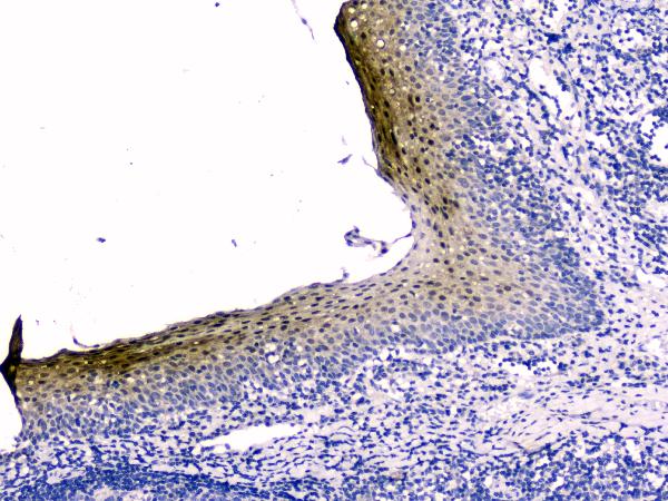
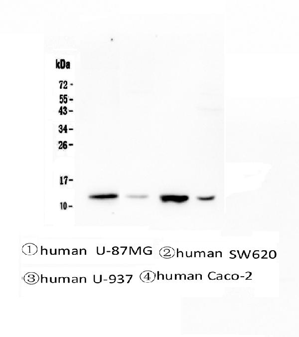
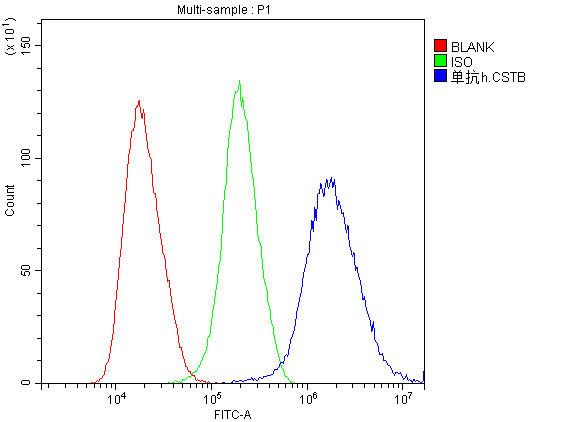
Anti-Stefin B CSTB Antibody Picoband™ (monoclonal, 2B6)
- SPECIFICATION
- CITATIONS
- PROTOCOLS
- BACKGROUND

Application
| WB, IHC, IHC-F, ICC, FC |
|---|---|
| Primary Accession | P04080 |
| Host | Mouse |
| Isotype | Mouse IgG1 |
| Reactivity | Human |
| Clonality | Monoclonal |
| Format | Lyophilized |
| Description | Anti-Stefin B CSTB Antibody Picoband™ (monoclonal, 2B6) . Tested in Flow Cytometry, IHC, IHC-F, ICC, WB applications. This antibody reacts with Human. |
| Reconstitution | Add 0.2ml of distilled water will yield a concentration of 500ug/ml. |
| Gene ID | 1476 |
|---|---|
| Other Names | Cystatin-B, CPI-B, Liver thiol proteinase inhibitor, Stefin-B, CSTB, CST6, STFB |
| Calculated MW | 14 kDa |
| Application Details | Western blot, 0.1-0.5 µg/ml Immunohistochemistry (Paraffin-embedded Section), 0.5-1 µg/ml Immunohistochemistry (Frozen Section), 0.5-1 µg/ml Immunocytochemistry, 0.5-1 µg/ml Flow Cytometry, 1-3 µg/1x10^6 cells |
| Subcellular Localization | Cytoplasm |
| Contents | Each vial contains 4mg Trehalose, 0.9mg NaCl, 0.2mg Na2HPO4, 0.05mg NaN3. |
| Clone Names | Clone: 2B6 |
| Immunogen | E. coli-derived human Stefin B recombinant protein (Position: M1-F98). Human Stefin B shares 78.6 % amino acid (aa) sequence identity with both mouse and rat Stefin B. |
| Cross Reactivity | No cross-reactivity with other proteins. |
| Storage | Store at -20˚C for one year from date of receipt. After reconstitution, at 4˚C for one month. It can also be aliquotted and stored frozen at -20˚C for six months. Avoid repeated freeze-thaw cycles. |
| Name | CSTB |
|---|---|
| Synonyms | CST6, STFB |
| Function | This is an intracellular thiol proteinase inhibitor. Tightly binding reversible inhibitor of cathepsins L, H and B. |
| Cellular Location | Cytoplasm. Nucleus |

Thousands of laboratories across the world have published research that depended on the performance of antibodies from Abcepta to advance their research. Check out links to articles that cite our products in major peer-reviewed journals, organized by research category.
info@abcepta.com, and receive a free "I Love Antibodies" mug.

Provided below are standard protocols that you may find useful for product applications.
Background
Cystatin B (CSTB), also called STFB, is a small protein that is a member of the superfamily of cysteine protease inhibitors. It has been isolated from human spleen and liver and its amino acid sequence has been fully determined. The cystatin B gene is located on 21q22.3. It is widely distributed and is localized mostly intracellularly, but has been found extracellularly. The protein is able to form a dimer stabilized by noncovalent forces, inhibiting papain and cathepsins l, h and b. Its role is thought to be as a protector against the proteinases leaking from lysosomes. A cystatin B multiprotein complex might have a specific cerebellar function, and that the loss of this function might contribute to the etiopathogenesis of EPM1. Upon differentiation to myotubes, CSTB becomes excluded from the nucleus and lysosomes, suggesting that the subcellular distribution of CSTB is dependent on the differentiation status of the cell.
If you have used an Abcepta product and would like to share how it has performed, please click on the "Submit Review" button and provide the requested information. Our staff will examine and post your review and contact you if needed.
If you have any additional inquiries please email technical services at tech@abcepta.com.
Ordering Information
Other Products
Shipping Information
