


 Foundational characteristics of cancer include proliferation, angiogenesis, migration, evasion of apoptosis, and cellular immortality. Find key markers for these cellular processes and antibodies to detect them.
 The SUMOplot™ Analysis Program predicts and scores sumoylation sites in your protein. SUMOylation is a post-translational modification involved in various cellular processes, such as nuclear-cytosolic transport, transcriptional regulation, apoptosis, protein stability, response to stress, and progression through the cell cycle.
 The Autophagy Receptor Motif Plotter predicts and scores autophagy receptor binding sites in your protein. Identifying proteins connected to this pathway is critical to understanding the role of autophagy in physiological as well as pathological processes such as development, differentiation, neurodegenerative diseases, stress, infection, and cancer.
Anti-APE1 APEX1 Antibody Picoband™ (monoclonal, 5C11)
- SPECIFICATION
- CITATIONS
- PROTOCOLS
- BACKGROUND

Application
| WB, IHC, ICC, FC |
|---|---|
| Primary Accession | P27695 |
| Host | Mouse |
| Isotype | Mouse IgG2b |
| Reactivity | Human |
| Clonality | Monoclonal |
| Format | Lyophilized |
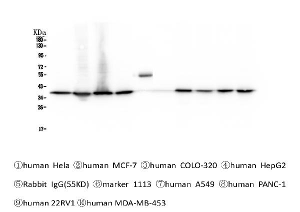
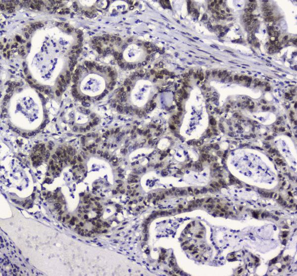
| Description | Anti-APE1 APEX1 Antibody Picoband™ (monoclonal, 5C11) . Tested in Flow Cytometry, IHC, ICC, WB applications. This antibody reacts with Human. |
| Reconstitution | Add 0.2ml of distilled water will yield a concentration of 500ug/ml. |
| Gene ID | 328 |
|---|---|
| Other Names | DNA repair nuclease/redox regulator APEX1, 3.1.11.2, 3.1.21.-, APEX nuclease, APEN, Apurinic-apyrimidinic endonuclease 1, AP endonuclease 1, APE-1, DNA-(apurinic or apyrimidinic site) endonuclease, Redox factor-1, REF-1, DNA repair nuclease/redox regulator APEX1, mitochondrial, APEX1, APE, APE1, APEX, APX, HAP1, REF1 |
| Calculated MW | 39 kDa |
| Application Details | Western blot, 0.1-0.5 µg/ml Immunohistochemistry (Paraffin-embedded Section), 0.5-1 µg/ml Immunohistochemistry (Frozen Section), 0.5-1 µg/ml Immunocytochemistry, 0.5-1 µg/ml Flow Cytometry, 1-3 µg/1x10^6 cells |
| Subcellular Localization | Nucleus. Nucleus, nucleolus. Nucleus speckle. Endoplasmic reticulum. Cytoplasm. Detected in the cytoplasm of B-cells stimulated to switch (By similarity). Colocalized with SIRT1 in the nucleus. Colocalized with YBX1 in nuclear speckles after genotoxic stress. Together with OGG1 is recruited to nuclear speckles in UVA-irradiated cells. Colocalized with nucleolin and NPM1 in the nucleolus. Its nucleolar localization is cell cycle dependent and requires active rRNA transcription. Colocalized with calreticulin in the endoplasmic reticulum. Translocation from the nucleus to the cytoplasm is stimulated in presence of nitric oxide (NO) and function in a CRM1-dependent manner, possibly as a consequence of demasking a nuclear export signal (amino acid position 64-80). S-nitrosylation at Cys-93 and Cys-310 regulates its nuclear-cytosolic shuttling. Ubiquitinated form is localized predominantly in the cytoplasm.. |
| Contents | Each vial contains 4mg Trehalose, 0.9mg NaCl, 0.2mg Na2HPO4, 0.05mg NaN3. |
| Clone Names | Clone: 5C11 |
| Immunogen | E.coli-derived human APE1 recombinant protein (Position: P2-L318). Human APE1 shares 94% and 93% amino acid (aa) sequence identity with mouse and rat APE1, respectively. |
| Cross Reactivity | No cross-reactivity with other proteins. |
| Storage | Store at -20˚C for one year from date of receipt. After reconstitution, at 4˚C for one month. It can also be aliquotted and stored frozen at -20˚C for six months. Avoid repeated freeze-thaw cycles. |
| Name | APEX1 |
|---|---|
| Synonyms | APE, APE1, APEX, APX, HAP1, REF1 |
| Function | Multifunctional protein that plays a central role in the cellular response to oxidative stress. The two major activities of APEX1 are DNA repair and redox regulation of transcriptional factors. Functions as an apurinic/apyrimidinic (AP) endodeoxyribonuclease in the DNA base excision repair (BER) pathway of DNA lesions induced by oxidative and alkylating agents. Initiates repair of AP sites in DNA by catalyzing hydrolytic incision of the phosphodiester backbone immediately adjacent to the damage, generating a single-strand break with 5'-deoxyribose phosphate and 3'-hydroxyl ends. Also incises at AP sites in the DNA strand of DNA/RNA hybrids, single-stranded DNA regions of R-loop structures, and single-stranded RNA molecules. Has 3'-5' exoribonuclease activity on mismatched deoxyribonucleotides at the 3' termini of nicked or gapped DNA molecules during short-patch BER. Possesses DNA 3' phosphodiesterase activity capable of removing lesions (such as phosphoglycolate) blocking the 3' side of DNA strand breaks. May also play a role in the epigenetic regulation of gene expression by participating in DNA demethylation. Acts as a loading factor for POLB onto non-incised AP sites in DNA and stimulates the 5'-terminal deoxyribose 5'-phosphate (dRp) excision activity of POLB. Plays a role in protection from granzyme-mediated cellular repair leading to cell death. Also involved in the DNA cleavage step of class switch recombination (CSR). On the other hand, APEX1 also exerts reversible nuclear redox activity to regulate DNA binding affinity and transcriptional activity of transcriptional factors by controlling the redox status of their DNA-binding domain, such as the FOS/JUN AP-1 complex after exposure to IR. Involved in calcium-dependent down- regulation of parathyroid hormone (PTH) expression by binding to negative calcium response elements (nCaREs). Together with HNRNPL or the dimer XRCC5/XRCC6, associates with nCaRE, acting as an activator of transcriptional repression. Stimulates the YBX1-mediated MDR1 promoter activity, when acetylated at Lys-6 and Lys-7, leading to drug resistance. Acts also as an endoribonuclease involved in the control of single-stranded RNA metabolism. Plays a role in regulating MYC mRNA turnover by preferentially cleaving in between UA and CA dinucleotides of the MYC coding region determinant (CRD). In association with NMD1, plays a role in the rRNA quality control process during cell cycle progression. Associates, together with YBX1, on the MDR1 promoter. Together with NPM1, associates with rRNA. Binds DNA and RNA. |
| Cellular Location | Nucleus {ECO:0000255|PROSITE-ProRule:PRU00764}. Nucleus, nucleolus. Nucleus speckle. Endoplasmic reticulum. Cytoplasm Note=Detected in the cytoplasm of B-cells stimulated to switch (By similarity). Colocalized with SIRT1 in the nucleus. Colocalized with YBX1 in nuclear speckles after genotoxic stress. Together with OGG1 is recruited to nuclear speckles in UVA-irradiated cells. Colocalized with nucleolin and NPM1 in the nucleolus. Its nucleolar localization is cell cycle dependent and requires active rRNA transcription. Colocalized with calreticulin in the endoplasmic reticulum. Translocation from the nucleus to the cytoplasm is stimulated in presence of nitric oxide (NO) and function in a CRM1-dependent manner, possibly as a consequence of demasking a nuclear export signal (amino acid position 64-80). S- nitrosylation at Cys-93 and Cys-310 regulates its nuclear-cytosolic shuttling. Ubiquitinated form is localized predominantly in the cytoplasm. |

Thousands of laboratories across the world have published research that depended on the performance of antibodies from Abcepta to advance their research. Check out links to articles that cite our products in major peer-reviewed journals, organized by research category.
info@abcepta.com, and receive a free "I Love Antibodies" mug.

Provided below are standard protocols that you may find useful for product applications.
Background
APEX1, also called apurinic endonuclease (APE), is a DNA repair enzyme having apurinic/apyrimidinic (AP) endonuclease, 3-prime, 5-prime-exonuclease, DNA 3-prime repair diesterase, and DNA 3-prime-phosphatase activities. The human APEX1 gene consists of 5 exons spanning 2.64 kb and exists as a single copy in the haploid genome. Using in situ hybridization, the APEX1 gene is mapped to 14q11.2-q12. The predicted APEX1 protein, which contained probable nuclear transport signals, was identified as a member of a family of DNA repair enzymes found in lower organisms. The abundance of the large form of APEX1 was increased in leiomyoma extracts relative to myometrial tissue extracts, and the large form was dominant in cell lines derived from leiomyosarcomas. The exonuclease activity of nuclear APEX1 can remove the anti-HIV nucleoside analogs AZT and D4T from the 3-prime terminus of a nick more efficiently than can cytosolic exonucleases.
If you have used an Abcepta product and would like to share how it has performed, please click on the "Submit Review" button and provide the requested information. Our staff will examine and post your review and contact you if needed.
If you have any additional inquiries please email technical services at tech@abcepta.com.
Ordering Information
Other Products
Shipping Information
