


 Foundational characteristics of cancer include proliferation, angiogenesis, migration, evasion of apoptosis, and cellular immortality. Find key markers for these cellular processes and antibodies to detect them.
 The SUMOplot™ Analysis Program predicts and scores sumoylation sites in your protein. SUMOylation is a post-translational modification involved in various cellular processes, such as nuclear-cytosolic transport, transcriptional regulation, apoptosis, protein stability, response to stress, and progression through the cell cycle.
 The Autophagy Receptor Motif Plotter predicts and scores autophagy receptor binding sites in your protein. Identifying proteins connected to this pathway is critical to understanding the role of autophagy in physiological as well as pathological processes such as development, differentiation, neurodegenerative diseases, stress, infection, and cancer.
> home > Products > Primary Antibodies > Signal Transduction > Anti-Calreticulin Rabbit Monoclonal Antibody
Anti-Calreticulin Rabbit Monoclonal Antibody
- SPECIFICATION
- CITATIONS
- PROTOCOLS
- BACKGROUND

Application
| WB, IHC, IF, ICC, IP, FC |
|---|---|
| Primary Accession | P27797 |
| Host | Rabbit |
| Isotype | Rabbit IgG |
| Reactivity | Rat, Human, Mouse |
| Clonality | Monoclonal |
| Format | Liquid |
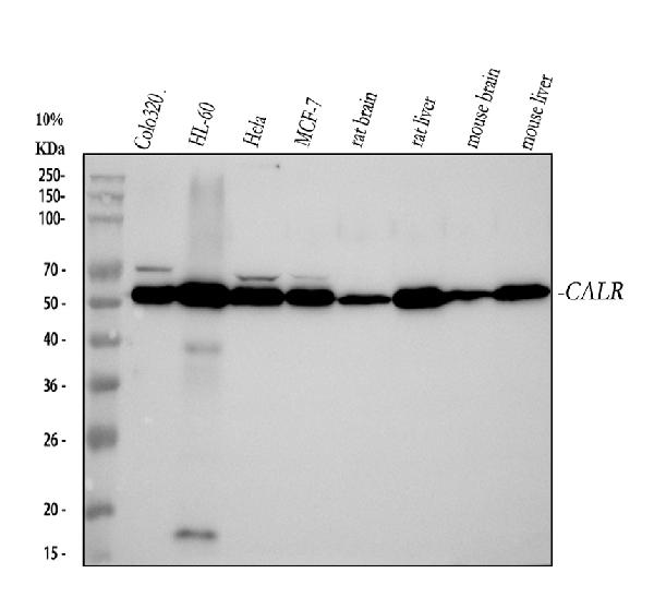
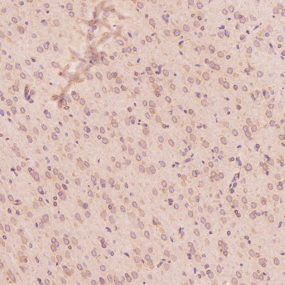
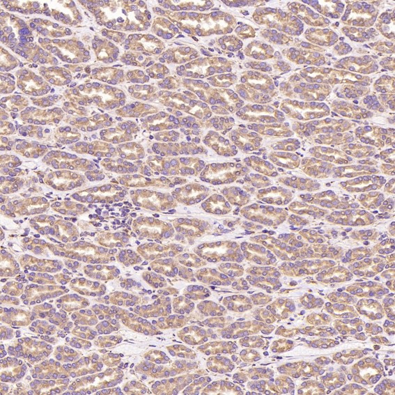
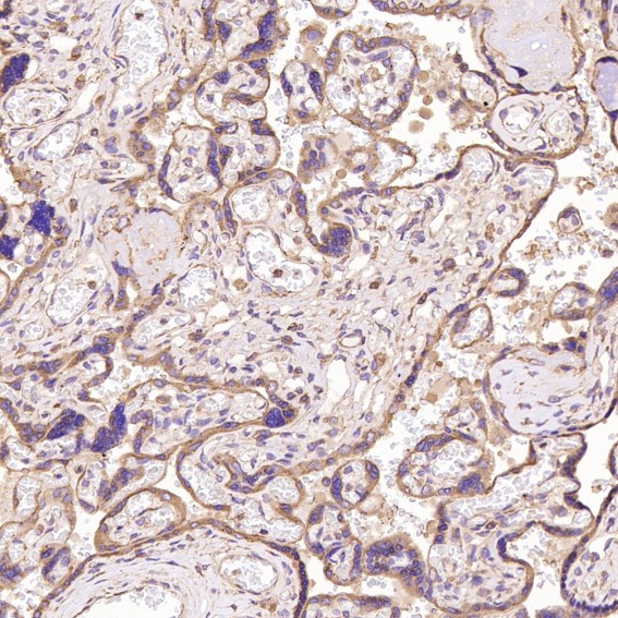
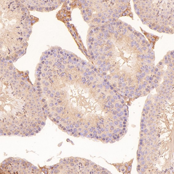
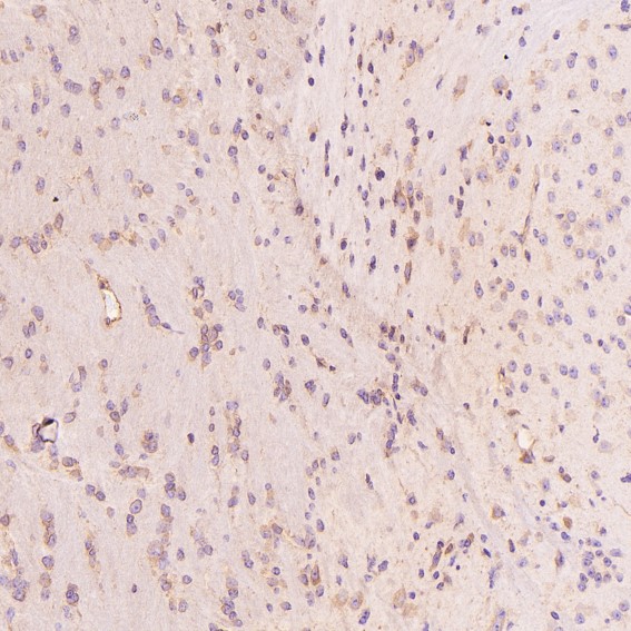
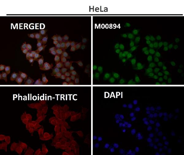
| Description | Anti-Calreticulin Rabbit Monoclonal Antibody . Tested in WB, IHC, ICC/IF, IP, Flow Cytometry applications. This antibody reacts with Human, Mouse, Rat. |
| Gene ID | 811 |
|---|---|
| Other Names | Calreticulin, CRP55, Calregulin, Endoplasmic reticulum resident protein 60, ERp60, HACBP, grp60, CALR (HGNC:1455), CRTC |
| Calculated MW | 48142 MW KDa |
| Application Details | WB 1:500-1:2000 IHC 1:50-1:200 ICC/IF 1:50-1:200 IP 1:50 FC 1:50 |
| Subcellular Localization | Endoplasmic reticulum lumen. Cytoplasm, cytosol. Secreted, extracellular space, extracellular matrix. Cell surface. Sarcoplasmic reticulum lumen. Also found in cell surface (T cells), cytosol and extracellular matrix (PubMed:10358038). Associated with the lytic granules in the cytolytic T-lymphocytes.. |
| Contents | Rabbit IgG in phosphate buffered saline, pH 7.4, 150mM NaCl, 0.02% sodium azide and 50% glycerol, 0.4-0.5mg/ml BSA. |
| Clone Names | Clone: CGO-3 |
| Immunogen | A synthesized peptide derived from human Calreticulin - ER Marker |
| Purification | Affinity-chromatography |
| Storage | Store at -20°C for one year. For short term storage and frequent use, store at 4°C for up to one month. Avoid repeated freeze-thaw cycles. |
| Name | CALR (HGNC:1455) |
|---|---|
| Synonyms | CRTC |
| Function | Calcium-binding chaperone that promotes folding, oligomeric assembly and quality control in the endoplasmic reticulum (ER) via the calreticulin/calnexin cycle. This lectin interacts transiently with almost all of the monoglucosylated glycoproteins that are synthesized in the ER (PubMed:7876246). Interacts with the DNA-binding domain of NR3C1 and mediates its nuclear export (PubMed:11149926). Involved in maternal gene expression regulation. May participate in oocyte maturation via the regulation of calcium homeostasis (By similarity). Present in the cortical granules of non-activated oocytes, is exocytosed during the cortical reaction in response to oocyte activation and might participate in the block to polyspermy (By similarity). |
| Cellular Location | Endoplasmic reticulum lumen. Cytoplasm, cytosol. Secreted, extracellular space, extracellular matrix. Cell surface. Sarcoplasmic reticulum lumen {ECO:0000250|UniProtKB:P28491}. Cytoplasmic vesicle, secretory vesicle, Cortical granule {ECO:0000250|UniProtKB:Q8K3H7}. Cytolytic granule. Note=Also found in cell surface (T cells), cytosol and extracellular matrix (PubMed:10358038). During oocyte maturation and after parthenogenetic activation accumulates in cortical granules. In pronuclear and early cleaved embryos localizes weakly to cytoplasm around nucleus and more strongly in the region near the cortex (By similarity). In cortical granules of non-activated oocytes, is exocytosed during the cortical reaction in response to oocyte activation (By similarity). {ECO:0000250|UniProtKB:P28491, ECO:0000250|UniProtKB:Q8K3H7, ECO:0000269|PubMed:8418194} |
Research Areas
Citations (0)

Thousands of laboratories across the world have published research that depended on the performance of antibodies from Abcepta to advance their research. Check out links to articles that cite our products in major peer-reviewed journals, organized by research category.
Submit your citation using an Abcepta antibody to
info@abcepta.com, and receive a free "I Love Antibodies" mug.
info@abcepta.com, and receive a free "I Love Antibodies" mug.

Application Protocols
Provided below are standard protocols that you may find useful for product applications.
Abcepta welcomes feedback from its customers.
If you have used an Abcepta product and would like to share how it has performed, please click on the "Submit Review" button and provide the requested information. Our staff will examine and post your review and contact you if needed.
If you have any additional inquiries please email technical services at tech@abcepta.com.
$ 370.00
Cat# ABO14120
Ordering Information
United States
AlbaniaAustraliaAustriaBelgiumBosnia & HerzegovinaBrazilBulgariaCanadaCentral AmericaChinaCroatiaCyprusCzech RepublicDenmarkEstoniaFinlandFranceGermanyGreeceHong KongHungaryIcelandIndiaIndonesiaIrelandIsraelItalyJapanLatviaLithuaniaLuxembourgMacedoniaMalaysiaMaltaNetherlandsNew ZealandNorwayPakistanPolandPortugalRomaniaSerbiaSingaporeSlovakiaSloveniaSouth AfricaSouth KoreaSpainSwedenSwitzerlandTaiwanTurkeyUnited KingdomUnited StatesVietnamWorldwideOthersMexico
USA Headquarters
(888) 735-7227 / (858) 622-0099 or (858) 875-1900
Other Products
Shipping Information
Domestic orders (in stock items)
Shipped out the same day. Orders placed after 1 PM (PST) will ship out the next business day.
International orders
Contact your local distributors
