


 Foundational characteristics of cancer include proliferation, angiogenesis, migration, evasion of apoptosis, and cellular immortality. Find key markers for these cellular processes and antibodies to detect them.
 The SUMOplot™ Analysis Program predicts and scores sumoylation sites in your protein. SUMOylation is a post-translational modification involved in various cellular processes, such as nuclear-cytosolic transport, transcriptional regulation, apoptosis, protein stability, response to stress, and progression through the cell cycle.
 The Autophagy Receptor Motif Plotter predicts and scores autophagy receptor binding sites in your protein. Identifying proteins connected to this pathway is critical to understanding the role of autophagy in physiological as well as pathological processes such as development, differentiation, neurodegenerative diseases, stress, infection, and cancer.
> home > Products > Primary Antibodies > Signal Transduction > Anti-Cathepsin B CTSB Rabbit Monoclonal Antibody
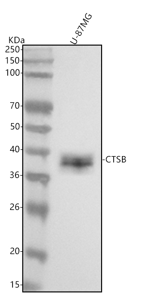
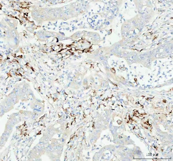
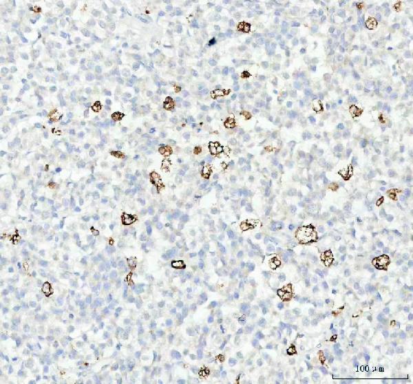
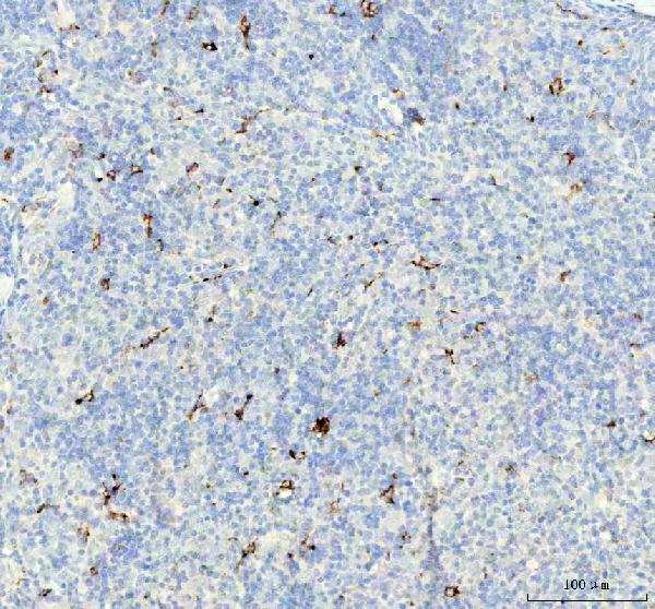
Anti-Cathepsin B CTSB Rabbit Monoclonal Antibody
- SPECIFICATION
- CITATIONS
- PROTOCOLS
- BACKGROUND

Application
| WB, IHC, IF, ICC, IP |
|---|---|
| Primary Accession | P07858 |
| Host | Rabbit |
| Isotype | Rabbit IgG |
| Reactivity | Human |
| Clonality | Monoclonal |
| Format | Liquid |
| Description | Anti-Cathepsin B CTSB Rabbit Monoclonal Antibody . Tested in WB, IHC, ICC/IF, IP applications. This antibody reacts with Human. |
| Gene ID | 1508 |
|---|---|
| Other Names | Cathepsin B, 3.4.22.1, APP secretase, APPS, Cathepsin B1, Cathepsin B light chain, Cathepsin B heavy chain, CTSB, CPSB |
| Calculated MW | 37822 MW KDa |
| Application Details | WB 1:500-1:2000 IHC 1:50-1:200 ICC/IF 1:50-1:200 IP 1:50 |
| Subcellular Localization | Lysosome. Melanosome. Secreted, extracellular space. Identified by mass spectrometry in melanosome fractions from stage I to stage IV. |
| Contents | Rabbit IgG in phosphate buffered saline, pH 7.4, 150mM NaCl, 0.02% sodium azide and 50% glycerol, 0.4-0.5mg/ml BSA. |
| Clone Names | Clone: AAFO-3 |
| Immunogen | A synthesized peptide derived from human Cathepsin B |
| Purification | Affinity-chromatography |
| Storage | Store at -20°C for one year. For short term storage and frequent use, store at 4°C for up to one month. Avoid repeated freeze-thaw cycles. |
| Name | CTSB |
|---|---|
| Synonyms | CPSB |
| Function | Thiol protease which is believed to participate in intracellular degradation and turnover of proteins (PubMed:12220505). Cleaves matrix extracellular phosphoglycoprotein MEPE (PubMed:12220505). Involved in the solubilization of cross-linked TG/thyroglobulin in the thyroid follicle lumen (By similarity). Has also been implicated in tumor invasion and metastasis (PubMed:3972105). |
| Cellular Location | Lysosome. Melanosome. Secreted, extracellular space {ECO:0000250|UniProtKB:A1E295}. Apical cell membrane {ECO:0000250|UniProtKB:P10605}; Peripheral membrane protein {ECO:0000250|UniProtKB:P10605}; Extracellular side {ECO:0000250|UniProtKB:P10605}. Note=Identified by mass spectrometry in melanosome fractions from stage I to stage IV (PubMed:17081065) Localizes to the lumen of thyroid follicles and to the apical membrane of thyroid epithelial cells (By similarity) {ECO:0000250|UniProtKB:P10605, ECO:0000269|PubMed:17081065} |
| Tissue Location | Expressed in the stratum spinosum of the epidermis. Weak expression is detected in the stratum granulosum |
Research Areas
Citations (0)

Thousands of laboratories across the world have published research that depended on the performance of antibodies from Abcepta to advance their research. Check out links to articles that cite our products in major peer-reviewed journals, organized by research category.
Submit your citation using an Abcepta antibody to
info@abcepta.com, and receive a free "I Love Antibodies" mug.
info@abcepta.com, and receive a free "I Love Antibodies" mug.

Application Protocols
Provided below are standard protocols that you may find useful for product applications.
Abcepta welcomes feedback from its customers.
If you have used an Abcepta product and would like to share how it has performed, please click on the "Submit Review" button and provide the requested information. Our staff will examine and post your review and contact you if needed.
If you have any additional inquiries please email technical services at tech@abcepta.com.
$ 370.00
Cat# ABO14105
Ordering Information
United States
AlbaniaAustraliaAustriaBelgiumBosnia & HerzegovinaBrazilBulgariaCanadaCentral AmericaChinaCroatiaCyprusCzech RepublicDenmarkEstoniaFinlandFranceGermanyGreeceHong KongHungaryIcelandIndiaIndonesiaIrelandIsraelItalyJapanLatviaLithuaniaLuxembourgMacedoniaMalaysiaMaltaNetherlandsNew ZealandNorwayPakistanPolandPortugalRomaniaSerbiaSingaporeSlovakiaSloveniaSouth AfricaSouth KoreaSpainSwedenSwitzerlandTaiwanTurkeyUnited KingdomUnited StatesVietnamWorldwideOthersMexico
USA Headquarters
(888) 735-7227 / (858) 622-0099 or (858) 875-1900
Other Products
Shipping Information
Domestic orders (in stock items)
Shipped out the same day. Orders placed after 1 PM (PST) will ship out the next business day.
International orders
Contact your local distributors
