


 Foundational characteristics of cancer include proliferation, angiogenesis, migration, evasion of apoptosis, and cellular immortality. Find key markers for these cellular processes and antibodies to detect them.
 The SUMOplot™ Analysis Program predicts and scores sumoylation sites in your protein. SUMOylation is a post-translational modification involved in various cellular processes, such as nuclear-cytosolic transport, transcriptional regulation, apoptosis, protein stability, response to stress, and progression through the cell cycle.
 The Autophagy Receptor Motif Plotter predicts and scores autophagy receptor binding sites in your protein. Identifying proteins connected to this pathway is critical to understanding the role of autophagy in physiological as well as pathological processes such as development, differentiation, neurodegenerative diseases, stress, infection, and cancer.
> home > Products > Primary Antibodies > Signal Transduction > Anti-Cortactin CTTN Rabbit Monoclonal Antibody
Anti-Cortactin CTTN Rabbit Monoclonal Antibody
- SPECIFICATION
- CITATIONS
- PROTOCOLS
- BACKGROUND

Application
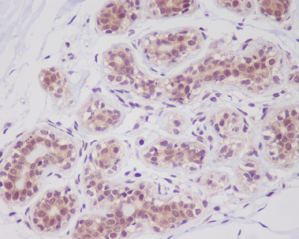
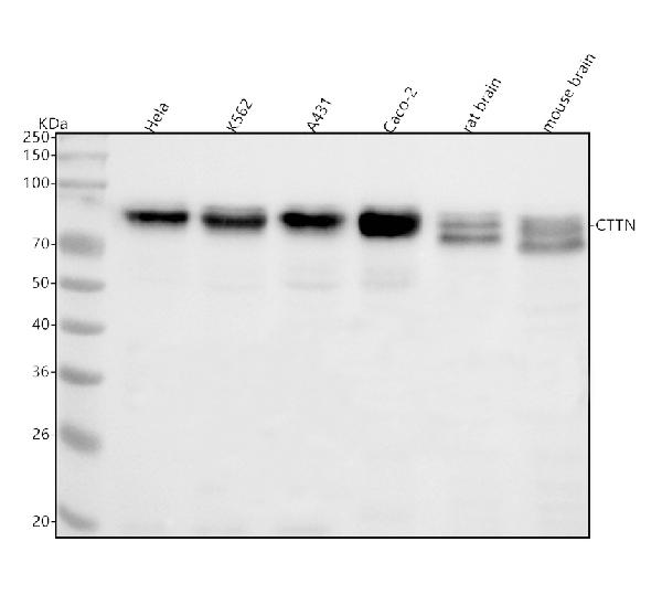
| WB, IHC, IF, ICC, IP, FC |
|---|---|
| Primary Accession | Q14247 |
| Host | Rabbit |
| Isotype | Rabbit IgG |
| Reactivity | Rat, Human, Mouse |
| Clonality | Monoclonal |
| Format | Liquid |
| Description | Anti-Cortactin CTTN Rabbit Monoclonal Antibody . Tested in WB, IHC, ICC/IF, IP, Flow Cytometry applications. This antibody reacts with Human, Mouse, Rat. |
| Gene ID | 2017 |
|---|---|
| Other Names | Src substrate cortactin, Amplaxin, Oncogene EMS1, CTTN, EMS1 |
| Calculated MW | 61586 MW KDa |
| Application Details | WB 1:5000-1:10000 IHC 1:50-1:200 ICC/IF 1:50-1:200 IP 1:20 FC 1:20 |
| Subcellular Localization | Cytoplasm, cytoskeleton. Cell projection, lamellipodium. Cell projection, ruffle. Cell projection, dendrite. Cell projection. Cell membrane ; Peripheral membrane protein ; Cytoplasmic side. Cell projection, podosome. Cell junction. Cell junction, focal adhesion. Membrane, clathrin-coated pit. Cell projection, dendritic spine. Cytoplasm, cell cortex. Colocalizes transiently with PTK2/FAK1 at focal adhesions (By similarity). Associated with membrane ruffles and lamellipodia. In the presence of CTTNBP2NL, colocalizes with stress fibers (By similarity). In the presence of CTTNBP2, localizes at the cell cortex (By similarity). In response to neuronal activation by glutamate, redistributes from dendritic spines to the dendritic shaft (By similarity). Colocalizes with DNM2 at the basis of filopodia in hippocampus neuron growth zones (By similarity).. |
| Contents | Rabbit IgG in phosphate buffered saline, pH 7.4, 150mM NaCl, 0.02% sodium azide and 50% glycerol, 0.4-0.5mg/ml BSA. |
| Clone Names | Clone: FAI-3 |
| Immunogen | A synthesized peptide derived from human Cortactin |
| Purification | Affinity-chromatography |
| Storage | Store at -20°C for one year. For short term storage and frequent use, store at 4°C for up to one month. Avoid repeated freeze-thaw cycles. |
| Name | CTTN |
|---|---|
| Synonyms | EMS1 |
| Function | Contributes to the organization of the actin cytoskeleton and cell shape (PubMed:21296879). Plays a role in the formation of lamellipodia and in cell migration. Plays a role in the regulation of neuron morphology, axon growth and formation of neuronal growth cones (By similarity). Through its interaction with CTTNBP2, involved in the regulation of neuronal spine density (By similarity). Plays a role in focal adhesion assembly and turnover (By similarity). In complex with ABL1 and MYLK regulates cortical actin-based cytoskeletal rearrangement critical to sphingosine 1-phosphate (S1P)-mediated endothelial cell (EC) barrier enhancement (PubMed:20861316). Plays a role in intracellular protein transport and endocytosis, and in modulating the levels of potassium channels present at the cell membrane (PubMed:17959782). Plays a role in receptor-mediated endocytosis via clathrin-coated pits (By similarity). Required for stabilization of KCNH1 channels at the cell membrane (PubMed:23144454). Plays a role in the invasiveness of cancer cells, and the formation of metastases (PubMed:16636290). |
| Cellular Location | Cytoplasm, cytoskeleton. Cell projection, lamellipodium. Cell projection, ruffle. Cell projection, dendrite. Cell projection {ECO:0000250|UniProtKB:Q66HL2}. Cell membrane; Peripheral membrane protein; Cytoplasmic side. Cell projection, podosome {ECO:0000250|UniProtKB:Q01406}. Cell junction {ECO:0000250|UniProtKB:Q66HL2}. Cell junction, focal adhesion {ECO:0000250|UniProtKB:Q66HL2}. Membrane, clathrin-coated pit {ECO:0000250|UniProtKB:Q66HL2}. Cell projection, dendritic spine. Cytoplasm, cell cortex Endoplasmic reticulum {ECO:0000250|UniProtKB:Q01406}. Note=Colocalizes transiently with PTK2/FAK1 at focal adhesions (By similarity) Associated with membrane ruffles and lamellipodia. In the presence of CTTNBP2NL, colocalizes with stress fibers (By similarity). In the presence of CTTNBP2, localizes at the cell cortex (By similarity). In response to neuronal activation by glutamate, redistributes from dendritic spines to the dendritic shaft (By similarity). Colocalizes with DNM2 at the basis of filopodia in hippocampus neuron growth zones (By similarity). {ECO:0000250|UniProtKB:Q60598, ECO:0000250|UniProtKB:Q66HL2} |
Research Areas
Citations (0)

Thousands of laboratories across the world have published research that depended on the performance of antibodies from Abcepta to advance their research. Check out links to articles that cite our products in major peer-reviewed journals, organized by research category.
Submit your citation using an Abcepta antibody to
info@abcepta.com, and receive a free "I Love Antibodies" mug.
info@abcepta.com, and receive a free "I Love Antibodies" mug.

Application Protocols
Provided below are standard protocols that you may find useful for product applications.
Abcepta welcomes feedback from its customers.
If you have used an Abcepta product and would like to share how it has performed, please click on the "Submit Review" button and provide the requested information. Our staff will examine and post your review and contact you if needed.
If you have any additional inquiries please email technical services at tech@abcepta.com.
$ 370.00
Cat# ABO13981
Ordering Information
United States
AlbaniaAustraliaAustriaBelgiumBosnia & HerzegovinaBrazilBulgariaCanadaCentral AmericaChinaCroatiaCyprusCzech RepublicDenmarkEstoniaFinlandFranceGermanyGreeceHong KongHungaryIcelandIndiaIndonesiaIrelandIsraelItalyJapanLatviaLithuaniaLuxembourgMacedoniaMalaysiaMaltaNetherlandsNew ZealandNorwayPakistanPolandPortugalRomaniaSerbiaSingaporeSlovakiaSloveniaSouth AfricaSouth KoreaSpainSwedenSwitzerlandTaiwanTurkeyUnited KingdomUnited StatesVietnamWorldwideOthersMexico
USA Headquarters
(888) 735-7227 / (858) 622-0099 or (858) 875-1900
Other Products
Shipping Information
Domestic orders (in stock items)
Shipped out the same day. Orders placed after 1 PM (PST) will ship out the next business day.
International orders
Contact your local distributors
