Anti-Filamin A FLNA Rabbit Monoclonal Antibody
- SPECIFICATION
- CITATIONS
- PROTOCOLS
- BACKGROUND

Application
| WB, IHC, IF, ICC, FC |
|---|---|
| Primary Accession | P21333 |
| Host | Rabbit |
| Isotype | Rabbit IgG |
| Reactivity | Rat, Human, Mouse |
| Clonality | Monoclonal |
| Format | Liquid |
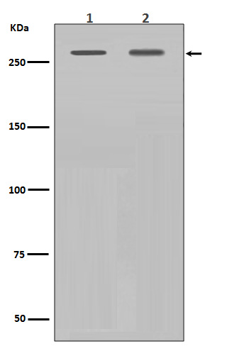
| Description | Anti-Filamin A FLNA Rabbit Monoclonal Antibody . Tested in WB, IHC, ICC/IF, Flow Cytometry applications. This antibody reacts with Human, Mouse, Rat. |
| Gene ID | 2316 |
|---|---|
| Other Names | Filamin-A, FLN-A, Actin-binding protein 280, ABP-280, Alpha-filamin, Endothelial actin-binding protein, Filamin-1, Non-muscle filamin, FLNA, FLN, FLN1 |
| Calculated MW | 280739 MW KDa |
| Application Details | WB 1:1000-1:2000 IHC 1:50-1:100 ICC/IF 1:50-1:100 FC 1:50 |
| Subcellular Localization | Cytoplasm, cell cortex. Cytoplasm, cytoskeleton. |
| Tissue Specificity | Ubiquitous. |
| Contents | Rabbit IgG in phosphate buffered saline, pH 7.4, 150mM NaCl, 0.02% sodium azide and 50% glycerol, 0.4-0.5mg/ml BSA. |
| Clone Names | Clone: AHF-6 |
| Immunogen | A synthesized peptide derived from human Filamin A |
| Purification | Affinity-chromatography |
| Storage | Store at -20°C for one year. For short term storage and frequent use, store at 4°C for up to one month. Avoid repeated freeze-thaw cycles. |
| Name | FLNA |
|---|---|
| Synonyms | FLN, FLN1 |
| Function | Promotes orthogonal branching of actin filaments and links actin filaments to membrane glycoproteins. Anchors various transmembrane proteins to the actin cytoskeleton and serves as a scaffold for a wide range of cytoplasmic signaling proteins. Interaction with FLNB may allow neuroblast migration from the ventricular zone into the cortical plate. Tethers cell surface- localized furin, modulates its rate of internalization and directs its intracellular trafficking (By similarity). Involved in ciliogenesis. Plays a role in cell-cell contacts and adherens junctions during the development of blood vessels, heart and brain organs. Plays a role in platelets morphology through interaction with SYK that regulates ITAM- and ITAM-like-containing receptor signaling, resulting in by platelet cytoskeleton organization maintenance (By similarity). During the axon guidance process, required for growth cone collapse induced by SEMA3A-mediated stimulation of neurons (PubMed:25358863). |
| Cellular Location | Cytoplasm, cell cortex. Cytoplasm, cytoskeleton {ECO:0000250|UniProtKB:Q8BTM8}. Perikaryon {ECO:0000250|UniProtKB:Q8BTM8}. Cell projection, growth cone {ECO:0000250|UniProtKB:Q8BTM8}. Cell projection, podosome {ECO:0000250|UniProtKB:Q8BTM8}. Note=Colocalizes with CPMR1 in the central region of DRG neuron growth cone (By similarity). Following SEMA3A stimulation of DRG neurons, colocalizes with F-actin (By similarity). Localized to the core of myotube podosomes (By similarity). {ECO:0000250|UniProtKB:Q8BTM8} |
| Tissue Location | Ubiquitous. |

Thousands of laboratories across the world have published research that depended on the performance of antibodies from Abcepta to advance their research. Check out links to articles that cite our products in major peer-reviewed journals, organized by research category.
info@abcepta.com, and receive a free "I Love Antibodies" mug.
Provided below are standard protocols that you may find useful for product applications.
If you have used an Abcepta product and would like to share how it has performed, please click on the "Submit Review" button and provide the requested information. Our staff will examine and post your review and contact you if needed.
If you have any additional inquiries please email technical services at tech@abcepta.com.


 Foundational characteristics of cancer include proliferation, angiogenesis, migration, evasion of apoptosis, and cellular immortality. Find key markers for these cellular processes and antibodies to detect them.
 The SUMOplot™ Analysis Program predicts and scores sumoylation sites in your protein. SUMOylation is a post-translational modification involved in various cellular processes, such as nuclear-cytosolic transport, transcriptional regulation, apoptosis, protein stability, response to stress, and progression through the cell cycle.
 The Autophagy Receptor Motif Plotter predicts and scores autophagy receptor binding sites in your protein. Identifying proteins connected to this pathway is critical to understanding the role of autophagy in physiological as well as pathological processes such as development, differentiation, neurodegenerative diseases, stress, infection, and cancer.


