Anti-NFAT2 NFATC1 Rabbit Monoclonal Antibody
- SPECIFICATION
- CITATIONS
- PROTOCOLS
- BACKGROUND

Application
| WB, IF, ICC, FC |
|---|---|
| Primary Accession | O95644 |
| Host | Rabbit |
| Isotype | Rabbit IgG |
| Reactivity | Human |
| Clonality | Monoclonal |
| Format | Liquid |
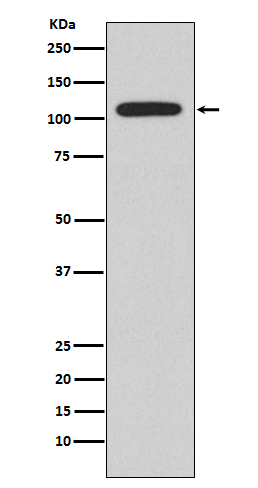
| Description | Anti-NFAT2 NFATC1 Rabbit Monoclonal Antibody . Tested in WB, ICC/IF, Flow Cytometry applications. This antibody reacts with Human. |
| Gene ID | 4772 |
|---|---|
| Other Names | Nuclear factor of activated T-cells, cytoplasmic 1, NF-ATc1, NFATc1, NFAT transcription complex cytosolic component, NF-ATc, NFATc, NFATC1, NFAT2, NFATC |
| Calculated MW | 101243 MW KDa |
| Application Details | WB 1:500-1:2000 ICC/IF 1:50-1:200 FC 1:50 |
| Subcellular Localization | Cytoplasm. Nucleus. Cytoplasmic for the phosphorylated form and nuclear after activation that is controlled by calcineurin-mediated dephosphorylation. Rapid nuclear exit of NFATC is thought to be one mechanism by which cells distinguish between sustained and transient calcium signals. The subcellular localization of NFATC plays a key role in the regulation of gene transcription. |
| Tissue Specificity | Expressed in thymus, peripheral leukocytes as T-cells and spleen. Isoforms A are preferentially expressed in effector T-cells (thymus and peripheral leukocytes) whereas isoforms B and isoforms C are preferentially expressed in naive T- cells (spleen). Isoforms B are expressed in naive T-cells after first antigen exposure and isoforms A are expressed in effector T- cells after second antigen exposure. Isoforms IA are widely expressed but not detected in liver nor pancreas, neural expression is strongest in corpus callosum. Isoforms IB are expressed mostly in muscle, cerebellum, placenta and thymus, neural expression in fetal and adult brain, strongest in corpus callosum.. |
| Contents | Rabbit IgG in phosphate buffered saline, pH 7.4, 150mM NaCl, 0.02% sodium azide and 50% glycerol, 0.4-0.5mg/ml BSA. |
| Clone Names | Clone: ABDA-14 |
| Immunogen | A synthesized peptide derived from human NFAT2 |
| Purification | Affinity-chromatography |
| Storage | Store at -20°C for one year. For short term storage and frequent use, store at 4°C for up to one month. Avoid repeated freeze-thaw cycles. |
| Name | NFATC1 |
|---|---|
| Synonyms | NFAT2, NFATC |
| Function | Plays a role in the inducible expression of cytokine genes in T-cells, especially in the induction of the IL-2 or IL-4 gene transcription. Also controls gene expression in embryonic cardiac cells. Could regulate not only the activation and proliferation but also the differentiation and programmed death of T-lymphocytes as well as lymphoid and non-lymphoid cells (PubMed:10358178). Required for osteoclastogenesis and regulates many genes important for osteoclast differentiation and function (By similarity). |
| Cellular Location | Cytoplasm. Nucleus. Note=Cytoplasmic for the phosphorylated form and nuclear after activation that is controlled by calcineurin- mediated dephosphorylation. Rapid nuclear exit of NFATC is thought to be one mechanism by which cells distinguish between sustained and transient calcium signals. Translocation to the nucleus is increased in the presence of calcium in pre-osteoblasts (By similarity). The subcellular localization of NFATC plays a key role in the regulation of gene transcription (PubMed:16511445). Nuclear translocation of NFATC1 is enhanced in the presence of TNFSF11. Nuclear translocation is decreased in the presence of FBN1 which can bind and sequester TNFSF11 (By similarity). {ECO:0000250|UniProtKB:O88942, ECO:0000269|PubMed:16511445} |
| Tissue Location | Expressed in thymus, peripheral leukocytes as T- cells and spleen. Isoforms A are preferentially expressed in effector T-cells (thymus and peripheral leukocytes) whereas isoforms B and isoforms C are preferentially expressed in naive T-cells (spleen) Isoforms B are expressed in naive T-cells after first antigen exposure and isoforms A are expressed in effector T-cells after second antigen exposure. Isoforms IA are widely expressed but not detected in liver nor pancreas, neural expression is strongest in corpus callosum Isoforms IB are expressed mostly in muscle, cerebellum, placenta and thymus, neural expression in fetal and adult brain, strongest in corpus callosum. |

Thousands of laboratories across the world have published research that depended on the performance of antibodies from Abcepta to advance their research. Check out links to articles that cite our products in major peer-reviewed journals, organized by research category.
info@abcepta.com, and receive a free "I Love Antibodies" mug.
Provided below are standard protocols that you may find useful for product applications.
If you have used an Abcepta product and would like to share how it has performed, please click on the "Submit Review" button and provide the requested information. Our staff will examine and post your review and contact you if needed.
If you have any additional inquiries please email technical services at tech@abcepta.com.


 Foundational characteristics of cancer include proliferation, angiogenesis, migration, evasion of apoptosis, and cellular immortality. Find key markers for these cellular processes and antibodies to detect them.
 The SUMOplot™ Analysis Program predicts and scores sumoylation sites in your protein. SUMOylation is a post-translational modification involved in various cellular processes, such as nuclear-cytosolic transport, transcriptional regulation, apoptosis, protein stability, response to stress, and progression through the cell cycle.
 The Autophagy Receptor Motif Plotter predicts and scores autophagy receptor binding sites in your protein. Identifying proteins connected to this pathway is critical to understanding the role of autophagy in physiological as well as pathological processes such as development, differentiation, neurodegenerative diseases, stress, infection, and cancer.


