


 Foundational characteristics of cancer include proliferation, angiogenesis, migration, evasion of apoptosis, and cellular immortality. Find key markers for these cellular processes and antibodies to detect them.
 The SUMOplot™ Analysis Program predicts and scores sumoylation sites in your protein. SUMOylation is a post-translational modification involved in various cellular processes, such as nuclear-cytosolic transport, transcriptional regulation, apoptosis, protein stability, response to stress, and progression through the cell cycle.
 The Autophagy Receptor Motif Plotter predicts and scores autophagy receptor binding sites in your protein. Identifying proteins connected to this pathway is critical to understanding the role of autophagy in physiological as well as pathological processes such as development, differentiation, neurodegenerative diseases, stress, infection, and cancer.
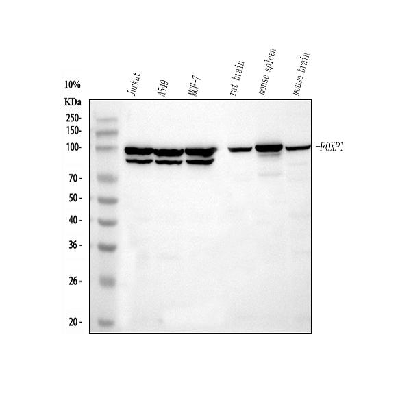
Anti-FOXP1 Rabbit Monoclonal Antibody
- SPECIFICATION
- CITATIONS
- PROTOCOLS
- BACKGROUND

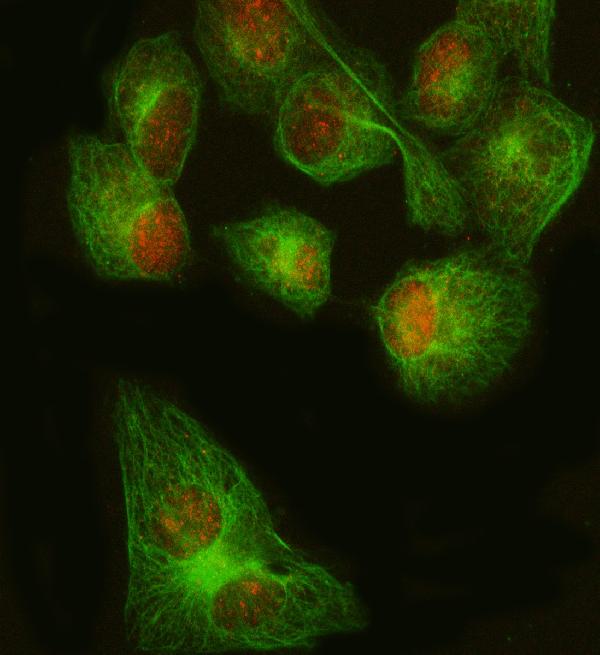
Application
| WB, IF, ICC |
|---|---|
| Primary Accession | Q9H334 |
| Host | Rabbit |
| Isotype | Rabbit IgG |
| Reactivity | Rat, Human, Mouse |
| Clonality | Monoclonal |
| Format | Liquid |
| Description | Anti-FOXP1 Rabbit Monoclonal Antibody . Tested in WB, ICC/IF applications. This antibody reacts with Human, Mouse, Rat. |
| Gene ID | 27086 |
|---|---|
| Other Names | Forkhead box protein P1, Mac-1-regulated forkhead, MFH, FOXP1 |
| Calculated MW | 75317 MW KDa |
| Application Details | WB 1:500-1:2000 ICC/IF 1:50-400 |
| Subcellular Localization | Nucleus. |
| Tissue Specificity | Isoform 8 is specifically expressed in embryonic stem cells.. |
| Contents | Rabbit IgG in phosphate buffered saline, pH 7.4, 150mM NaCl, 0.02% sodium azide and 50% glycerol, 0.4-0.5mg/ml BSA. |
| Clone Names | Clone: AOGO-6 |
| Immunogen | A synthesized peptide derived from human FOXP1 |
| Purification | Affinity-chromatography |
| Storage | Store at -20°C for one year. For short term storage and frequent use, store at 4°C for up to one month. Avoid repeated freeze-thaw cycles. |
| Name | FOXP1 |
|---|---|
| Function | Transcriptional repressor (PubMed:18347093, PubMed:26647308). Can act with CTBP1 to synergistically repress transcription but CTPBP1 is not essential (By similarity). Plays an important role in the specification and differentiation of lung epithelium. Acts cooperatively with FOXP4 to regulate lung secretory epithelial cell fate and regeneration by restricting the goblet cell lineage program; the function may involve regulation of AGR2. Essential transcriptional regulator of B-cell development. Involved in regulation of cardiac muscle cell proliferation. Involved in the columnar organization of spinal motor neurons. Promotes the formation of the lateral motor neuron column (LMC) and the preganglionic motor column (PGC) and is required for respective appropriate motor axon projections. The segment-appropriate generation of spinal cord motor columns requires cooperation with other Hox proteins. Can regulate PITX3 promoter activity; may promote midbrain identity in embryonic stem cell-derived dopamine neurons by regulating PITX3. Negatively regulates the differentiation of T follicular helper cells T(FH)s. Involved in maintenance of hair follicle stem cell quiescence; the function probably involves regulation of FGF18 (By similarity). Represses transcription of various pro-apoptotic genes and cooperates with NF- kappa B-signaling in promoting B-cell expansion by inhibition of caspase-dependent apoptosis (PubMed:25267198). Binds to CSF1R promoter elements and is involved in regulation of monocyte differentiation and macrophage functions; repression of CSF1R in monocytes seems to involve NCOR2 as corepressor (PubMed:15286807, PubMed:18347093, PubMed:18799727). Involved in endothelial cell proliferation, tube formation and migration indicative for a role in angiogenesis; the role in neovascularization seems to implicate suppression of SEMA5B (PubMed:24023716). Can negatively regulate androgen receptor signaling (PubMed:18640093). Acts as a transcriptional activator of the FBXL7 promoter; this activity is regulated by AURKA (PubMed:28218735). |
| Cellular Location | Nucleus. Note=Not found in the nucleolus |
| Tissue Location | Isoform 8 is specifically expressed in embryonic stem cells. |
Research Areas
Citations (0)

Thousands of laboratories across the world have published research that depended on the performance of antibodies from Abcepta to advance their research. Check out links to articles that cite our products in major peer-reviewed journals, organized by research category.
Submit your citation using an Abcepta antibody to
info@abcepta.com, and receive a free "I Love Antibodies" mug.
info@abcepta.com, and receive a free "I Love Antibodies" mug.

Application Protocols
Provided below are standard protocols that you may find useful for product applications.
Abcepta welcomes feedback from its customers.
If you have used an Abcepta product and would like to share how it has performed, please click on the "Submit Review" button and provide the requested information. Our staff will examine and post your review and contact you if needed.
If you have any additional inquiries please email technical services at tech@abcepta.com.
$ 370.00
Cat# ABO13747
Ordering Information
United States
AlbaniaAustraliaAustriaBelgiumBosnia & HerzegovinaBrazilBulgariaCanadaCentral AmericaChinaCroatiaCyprusCzech RepublicDenmarkEstoniaFinlandFranceGermanyGreeceHong KongHungaryIcelandIndiaIndonesiaIrelandIsraelItalyJapanLatviaLithuaniaLuxembourgMacedoniaMalaysiaMaltaNetherlandsNew ZealandNorwayPakistanPolandPortugalRomaniaSerbiaSingaporeSlovakiaSloveniaSouth AfricaSouth KoreaSpainSwedenSwitzerlandTaiwanTurkeyUnited KingdomUnited StatesVietnamWorldwideOthersMexico
USA Headquarters
(888) 735-7227 / (858) 622-0099 or (858) 875-1900
Other Products
Shipping Information
Domestic orders (in stock items)
Shipped out the same day. Orders placed after 1 PM (PST) will ship out the next business day.
International orders
Contact your local distributors
