


 Foundational characteristics of cancer include proliferation, angiogenesis, migration, evasion of apoptosis, and cellular immortality. Find key markers for these cellular processes and antibodies to detect them.
 The SUMOplot™ Analysis Program predicts and scores sumoylation sites in your protein. SUMOylation is a post-translational modification involved in various cellular processes, such as nuclear-cytosolic transport, transcriptional regulation, apoptosis, protein stability, response to stress, and progression through the cell cycle.
 The Autophagy Receptor Motif Plotter predicts and scores autophagy receptor binding sites in your protein. Identifying proteins connected to this pathway is critical to understanding the role of autophagy in physiological as well as pathological processes such as development, differentiation, neurodegenerative diseases, stress, infection, and cancer.
> home > Products > Primary Antibodies > Signal Transduction > Anti-CDC42 Rabbit Monoclonal Antibody
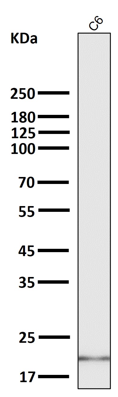
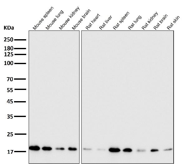
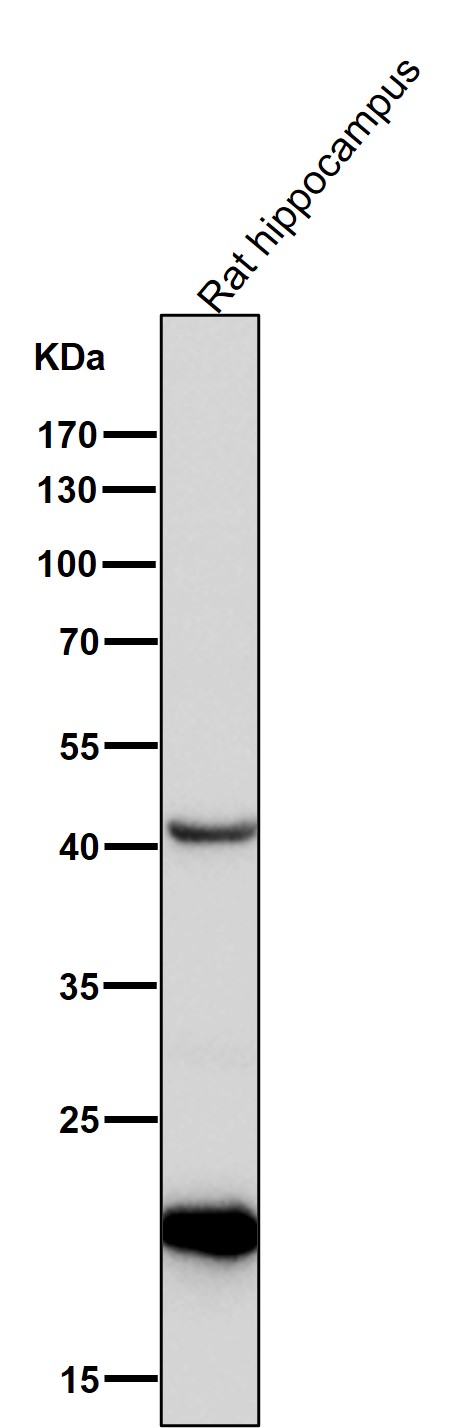
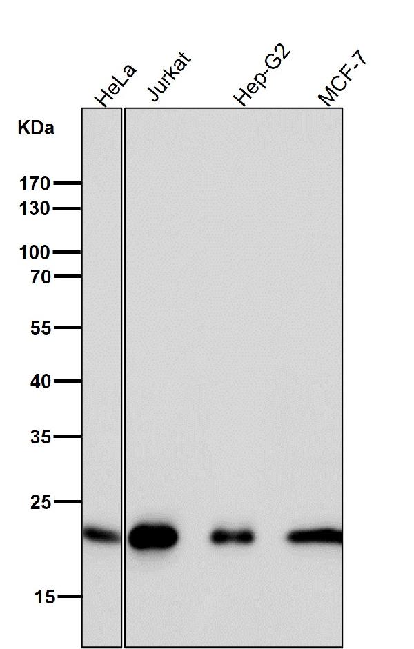
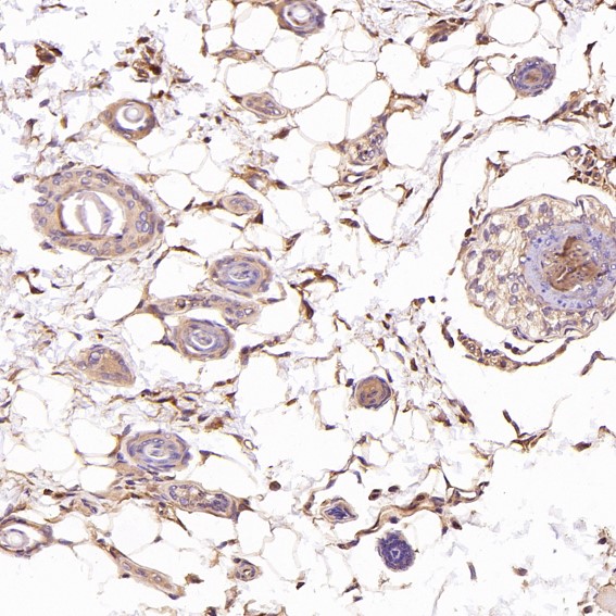
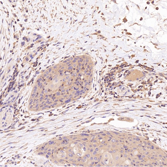
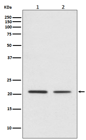
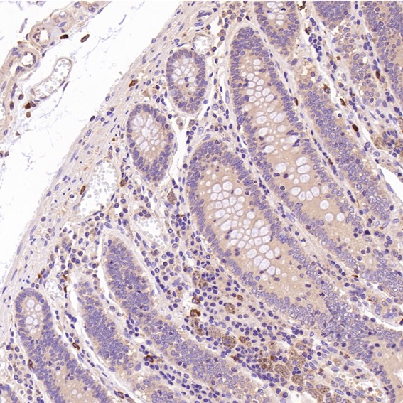
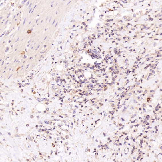
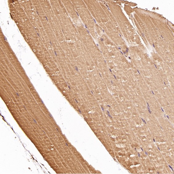
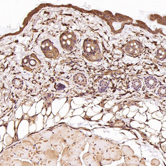
Anti-CDC42 Rabbit Monoclonal Antibody
- SPECIFICATION
- CITATIONS
- PROTOCOLS
- BACKGROUND

Application
| WB, IHC, IP, FC |
|---|---|
| Primary Accession | P60953 |
| Host | Rabbit |
| Isotype | Rabbit IgG |
| Reactivity | Rat, Human, Mouse |
| Clonality | Monoclonal |
| Format | Liquid |
| Description | Anti-CDC42 Rabbit Monoclonal Antibody . Tested in WB, IHC, IP, Flow Cytometry applications. This antibody reacts with Human, Mouse, Rat. |
| Gene ID | 998 |
|---|---|
| Other Names | Cell division control protein 42 homolog, 3.6.5.2, G25K GTP-binding protein, CDC42 (HGNC:1736) |
| Calculated MW | 21259 MW KDa |
| Application Details | WB 1:1000-1:2000 IHC 1:50-1:200 IP 1:30 FC 1:50 |
| Subcellular Localization | Cell membrane ; Lipid-anchor ; Cytoplasmic side. Cytoplasm, cytoskeleton, microtubule organizing center, centrosome. Cytoplasm, cytoskeleton, spindle. Midbody. Localizes to spindle during prometaphase cells. Moves to the central spindle as cells progressed through anaphase to telophase. Localizes at the end of cytokinesis in the intercellular bridge formed between two daughter cells. Its localization is regulated by the activities of guanine nucleotide exchange factor ECT2 and GTPase activating protein RACGAP1. Colocalizes with NEK6 in the centrosome. |
| Contents | Rabbit IgG in phosphate buffered saline, pH 7.4, 150mM NaCl, 0.02% sodium azide and 50% glycerol, 0.4-0.5mg/ml BSA. |
| Clone Names | Clone: IAE-3 |
| Immunogen | A synthesized peptide derived from human CDC42 |
| Purification | Affinity-chromatography |
| Storage | Store at -20°C for one year. For short term storage and frequent use, store at 4°C for up to one month. Avoid repeated freeze-thaw cycles. |
| Name | CDC42 (HGNC:1736) |
|---|---|
| Function | Plasma membrane-associated small GTPase which cycles between an active GTP-bound and an inactive GDP-bound state. In active state binds to a variety of effector proteins to regulate cellular responses. Involved in epithelial cell polarization processes. Regulates the bipolar attachment of spindle microtubules to kinetochores before chromosome congression in metaphase (PubMed:15642749). Regulates cell migration (PubMed:17038317, PubMed:22843693). In neurons, plays a role in the extension and maintenance of the formation of filopodia, thin and actin-rich surface projections (PubMed:14978216). Required for DOCK10-mediated spine formation in Purkinje cells and hippocampal neurons. In podocytes, facilitates filopodia and podosomes formation upon DOCK11-activation (PubMed:33523862). Upon activation by CaMKII, modulates dendritic spine structural plasticity by relaying CaMKII transient activation to synapse-specific, long-term signaling (By similarity). Also plays a role in phagocytosis through organization of the F-actin cytoskeleton associated with forming phagocytic cups (PubMed:26465210). Upon activation by PLEKHG4B, involved in actin cytoskeletal remodeling during epithelial cell-cell junction formation (PubMed:33310911). |
| Cellular Location | Cell membrane; Lipid-anchor; Cytoplasmic side. Cytoplasm, cytoskeleton, microtubule organizing center, centrosome. Cytoplasm, cytoskeleton, spindle. Midbody Cell projection, dendrite {ECO:0000250|UniProtKB:P60766} Note=Localizes to spindle during prometaphase cells. Moves to the central spindle as cells progressed through anaphase to telophase (PubMed:15642749). Localizes at the end of cytokinesis in the intercellular bridge formed between two daughter cells (PubMed:15642749). Its localization is regulated by the activities of guanine nucleotide exchange factor ECT2 and GTPase activating protein RACGAP1 (PubMed:15642749). Colocalizes with NEK6 in the centrosome (PubMed:20873783). In its active GTP-bound form localizes to the leading edge membrane of migrating dendritic cells (By similarity) {ECO:0000250|UniProtKB:P60766, ECO:0000269|PubMed:15642749, ECO:0000269|PubMed:20873783} |
Research Areas
Citations (0)

Thousands of laboratories across the world have published research that depended on the performance of antibodies from Abcepta to advance their research. Check out links to articles that cite our products in major peer-reviewed journals, organized by research category.
Submit your citation using an Abcepta antibody to
info@abcepta.com, and receive a free "I Love Antibodies" mug.
info@abcepta.com, and receive a free "I Love Antibodies" mug.

Application Protocols
Provided below are standard protocols that you may find useful for product applications.
Abcepta welcomes feedback from its customers.
If you have used an Abcepta product and would like to share how it has performed, please click on the "Submit Review" button and provide the requested information. Our staff will examine and post your review and contact you if needed.
If you have any additional inquiries please email technical services at tech@abcepta.com.
$ 370.00
Cat# ABO13712
Ordering Information
United States
AlbaniaAustraliaAustriaBelgiumBosnia & HerzegovinaBrazilBulgariaCanadaCentral AmericaChinaCroatiaCyprusCzech RepublicDenmarkEstoniaFinlandFranceGermanyGreeceHong KongHungaryIcelandIndiaIndonesiaIrelandIsraelItalyJapanLatviaLithuaniaLuxembourgMacedoniaMalaysiaMaltaNetherlandsNew ZealandNorwayPakistanPolandPortugalRomaniaSerbiaSingaporeSlovakiaSloveniaSouth AfricaSouth KoreaSpainSwedenSwitzerlandTaiwanTurkeyUnited KingdomUnited StatesVietnamWorldwideOthers
USA Headquarters
(888) 735-7227 / (858) 622-0099 or (858) 875-1900
Other Products
Shipping Information
Domestic orders (in stock items)
Shipped out the same day. Orders placed after 1 PM (PST) will ship out the next business day.
International orders
Contact your local distributors
