


 Foundational characteristics of cancer include proliferation, angiogenesis, migration, evasion of apoptosis, and cellular immortality. Find key markers for these cellular processes and antibodies to detect them.
 The SUMOplot™ Analysis Program predicts and scores sumoylation sites in your protein. SUMOylation is a post-translational modification involved in various cellular processes, such as nuclear-cytosolic transport, transcriptional regulation, apoptosis, protein stability, response to stress, and progression through the cell cycle.
 The Autophagy Receptor Motif Plotter predicts and scores autophagy receptor binding sites in your protein. Identifying proteins connected to this pathway is critical to understanding the role of autophagy in physiological as well as pathological processes such as development, differentiation, neurodegenerative diseases, stress, infection, and cancer.
> home > Products > Primary Antibodies > Signal Transduction > Anti-ROCK1 Rabbit Monoclonal Antibody
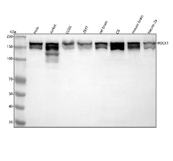
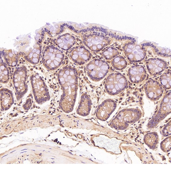
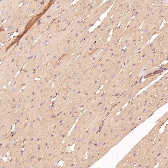
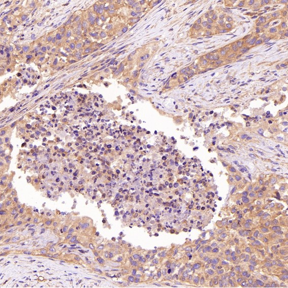
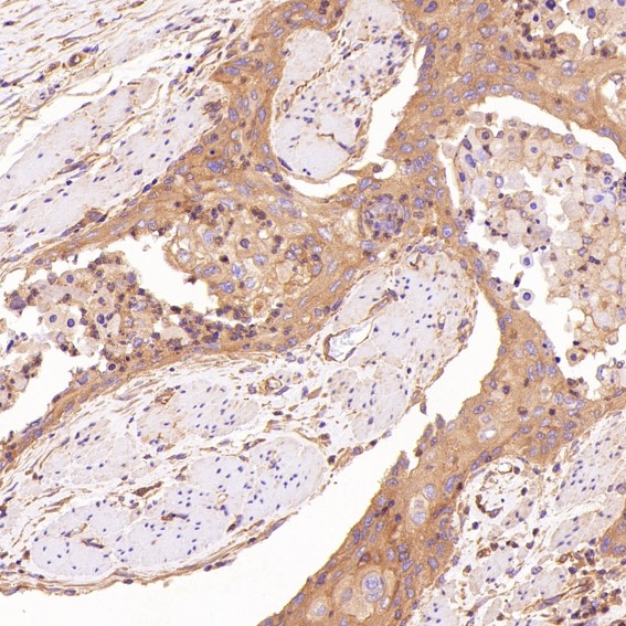
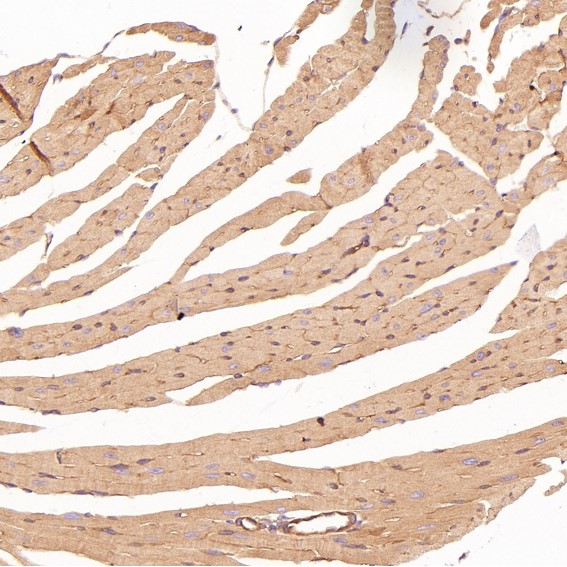
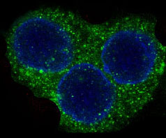
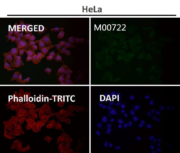
Anti-ROCK1 Rabbit Monoclonal Antibody
- SPECIFICATION
- CITATIONS
- PROTOCOLS
- BACKGROUND

Application
| WB, IHC, IF, ICC, IP, FC |
|---|---|
| Primary Accession | Q13464 |
| Host | Rabbit |
| Isotype | Rabbit IgG |
| Reactivity | Rat, Human, Mouse |
| Clonality | Monoclonal |
| Format | Liquid |
| Description | Anti-ROCK1 Rabbit Monoclonal Antibody . Tested in WB, IHC, ICC/IF, IP, Flow Cytometry applications. This antibody reacts with Human, Mouse, Rat. |
| Gene ID | 6093 |
|---|---|
| Other Names | Rho-associated protein kinase 1, 2.7.11.1, Renal carcinoma antigen NY-REN-35, Rho-associated, coiled-coil-containing protein kinase 1, Rho-associated, coiled-coil-containing protein kinase I, ROCK-I, p160 ROCK-1, p160ROCK, ROCK1 |
| Calculated MW | 158175 MW KDa |
| Application Details | WB 1:500-1:2000 IHC 1:50-1:200 ICC/IF 1:50-1:200 IP 1:50 FC 1:50 |
| Subcellular Localization | Cytoplasm. Cytoplasm, cytoskeleton, microtubule organizing center, centrosome, centriole. Golgi apparatus membrane; Peripheral membrane protein. Cell projection, bleb. Cytoplasm, cytoskeleton. Cell membrane. Cell projection, lamellipodium. Cell projection, ruffle. Associated with the mother centriole and an intercentriolar linker. Colocalizes with ITGB1BP1 and ITGB1 at the cell membrane predominantly in lamellipodia and membrane ruffles, but also in retraction fibers. Localizes at the cell membrane in an ITGB1BP1-dependent manner (By similarity). A small proportion is associated with Golgi membranes.. |
| Tissue Specificity | Detected in blood platelets.. |
| Contents | Rabbit IgG in phosphate buffered saline, pH 7.4, 150mM NaCl, 0.02% sodium azide and 50% glycerol, 0.4-0.5mg/ml BSA. |
| Clone Names | Clone: CDE-18 |
| Immunogen | A synthesized peptide derived from human ROCK1 |
| Purification | Affinity-chromatography |
| Storage | Store at -20°C for one year. For short term storage and frequent use, store at 4°C for up to one month. Avoid repeated freeze-thaw cycles. |
| Name | ROCK1 |
|---|---|
| Function | Protein kinase which is a key regulator of the actin cytoskeleton and cell polarity (PubMed:10436159, PubMed:10652353, PubMed:11018042, PubMed:11283607, PubMed:17158456, PubMed:18573880, PubMed:19131646, PubMed:8617235, PubMed:9722579). Involved in regulation of smooth muscle contraction, actin cytoskeleton organization, stress fiber and focal adhesion formation, neurite retraction, cell adhesion and motility via phosphorylation of DAPK3, GFAP, LIMK1, LIMK2, MYL9/MLC2, TPPP, PFN1 and PPP1R12A (PubMed:10436159, PubMed:10652353, PubMed:11018042, PubMed:11283607, PubMed:17158456, PubMed:18573880, PubMed:19131646, PubMed:23093407, PubMed:23355470, PubMed:8617235, PubMed:9722579). Phosphorylates FHOD1 and acts synergistically with it to promote SRC-dependent non-apoptotic plasma membrane blebbing (PubMed:18694941). Phosphorylates JIP3 and regulates the recruitment of JNK to JIP3 upon UVB-induced stress (PubMed:19036714). Acts as a suppressor of inflammatory cell migration by regulating PTEN phosphorylation and stability (By similarity). Acts as a negative regulator of VEGF-induced angiogenic endothelial cell activation (PubMed:19181962). Required for centrosome positioning and centrosome-dependent exit from mitosis (By similarity). Plays a role in terminal erythroid differentiation (PubMed:21072057). Inhibits podocyte motility via regulation of actin cytoskeletal dynamics and phosphorylation of CFL1 (By similarity). Promotes keratinocyte terminal differentiation (PubMed:19997641). Involved in osteoblast compaction through the fibronectin fibrillogenesis cell-mediated matrix assembly process, essential for osteoblast mineralization (By similarity). May regulate closure of the eyelids and ventral body wall by inducing the assembly of actomyosin bundles (By similarity). |
| Cellular Location | Cytoplasm. Cytoplasm, cytoskeleton, microtubule organizing center, centrosome, centriole {ECO:0000250|UniProtKB:P70335}. Golgi apparatus membrane; Peripheral membrane protein. Cell projection, bleb. Cytoplasm, cytoskeleton {ECO:0000250|UniProtKB:P70335}. Cell membrane {ECO:0000250|UniProtKB:P70335}. Cell projection, lamellipodium {ECO:0000250|UniProtKB:P70335}. Cell projection, ruffle {ECO:0000250|UniProtKB:P70335}. Note=A small proportion is associated with Golgi membranes (PubMed:12773565). Associated with the mother centriole and an intercentriolar linker (By similarity). Colocalizes with ITGB1BP1 and ITGB1 at the cell membrane predominantly in lamellipodia and membrane ruffles, but also in retraction fibers (By similarity). Localizes at the cell membrane in an ITGB1BP1-dependent manner (By similarity). {ECO:0000250|UniProtKB:P70335, ECO:0000269|PubMed:12773565} |
| Tissue Location | Detected in blood platelets. |
Research Areas
Citations (0)

Thousands of laboratories across the world have published research that depended on the performance of antibodies from Abcepta to advance their research. Check out links to articles that cite our products in major peer-reviewed journals, organized by research category.
Submit your citation using an Abcepta antibody to
info@abcepta.com, and receive a free "I Love Antibodies" mug.
info@abcepta.com, and receive a free "I Love Antibodies" mug.

Application Protocols
Provided below are standard protocols that you may find useful for product applications.
Abcepta welcomes feedback from its customers.
If you have used an Abcepta product and would like to share how it has performed, please click on the "Submit Review" button and provide the requested information. Our staff will examine and post your review and contact you if needed.
If you have any additional inquiries please email technical services at tech@abcepta.com.
$ 370.00
Cat# ABO13624
Ordering Information
United States
AlbaniaAustraliaAustriaBelgiumBosnia & HerzegovinaBrazilBulgariaCanadaCentral AmericaChinaCroatiaCyprusCzech RepublicDenmarkEstoniaFinlandFranceGermanyGreeceHong KongHungaryIcelandIndiaIndonesiaIrelandIsraelItalyJapanLatviaLithuaniaLuxembourgMacedoniaMalaysiaMaltaNetherlandsNew ZealandNorwayPakistanPolandPortugalRomaniaSerbiaSingaporeSlovakiaSloveniaSouth AfricaSouth KoreaSpainSwedenSwitzerlandTaiwanTurkeyUnited KingdomUnited StatesVietnamWorldwideOthers
USA Headquarters
(888) 735-7227 / (858) 622-0099 or (858) 875-1900
Other Products
Shipping Information
Domestic orders (in stock items)
Shipped out the same day. Orders placed after 1 PM (PST) will ship out the next business day.
International orders
Contact your local distributors
