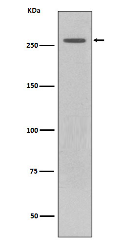
Anti-LRRK2 Rabbit Monoclonal Antibody
- SPECIFICATION
- CITATIONS
- PROTOCOLS
- BACKGROUND

Application
| WB, IHC, IF, ICC |
|---|---|
| Primary Accession | Q5S007 |
| Host | Rabbit |
| Isotype | Rabbit IgG |
| Reactivity | Human, Mouse |
| Clonality | Monoclonal |
| Format | Liquid |
| Description | Anti-LRRK2 Rabbit Monoclonal Antibody . Tested in WB, IHC, ICC/IF applications. This antibody reacts with Human, Mouse. |
| Gene ID | 120892 |
|---|---|
| Other Names | Leucine-rich repeat serine/threonine-protein kinase 2, 2.7.11.1, 3.6.5.-, Dardarin, LRRK2, PARK8 |
| Calculated MW | 286103 MW KDa |
| Application Details | WB 1:500-1:2000 IHC 1:50-1:200 ICC/IF 1:50-1:200 |
| Subcellular Localization | Membrane; Peripheral membrane protein. Cytoplasm. Perikaryon. Mitochondrion. Golgi apparatus. Cell projection, axon. Cell projection, dendrite. Endoplasmic reticulum. Cytoplasmic vesicle, secretory vesicle, synaptic vesicle membrane ; Peripheral membrane protein ; Cytoplasmic side. Endosome. Lysosome. Mitochondrion outer membrane. Mitochondrion inner membrane. Mitochondrion matrix. Predominantly associated with intracytoplasmic vesicular and membranous structures (By similarity). Localized in the cytoplasm and associated with cellular membrane structures. Predominantly associated with the mitochondrial outer membrane of the mitochondria. Colocalized with RAB29 along tubular structures emerging from Golgi apparatus. Localizes in intracytoplasmic punctate structures of neuronal perikarya and dendritic and axonal processes.. |
| Tissue Specificity | Expressed in the brain. Expressed in pyramidal neurons in all cortical laminae of the visual cortex, in neurons of the substantia nigra pars compacta and caudate putamen (at protein level). Expressed throughout the adult brain, but at a lower level than in heart and liver. Also expressed in placenta, lung, skeletal muscle, kidney and pancreas. In the brain, expressed in the cerebellum, cerebral cortex, medulla, spinal cord occipital pole, frontal lobe, temporal lobe and putamen. Expression is particularly high in brain dopaminoceptive areas.. |
| Contents | Rabbit IgG in phosphate buffered saline, pH 7.4, 150mM NaCl, 0.02% sodium azide and 50% glycerol, 0.4-0.5mg/ml BSA. |
| Clone Names | Clone: BBG-12 |
| Immunogen | A synthesized peptide derived from human LRRK2 |
| Purification | Affinity-chromatography |
| Storage | Store at -20°C for one year. For short term storage and frequent use, store at 4°C for up to one month. Avoid repeated freeze-thaw cycles. |
| Name | LRRK2 |
|---|---|
| Synonyms | PARK8 |
| Function | Serine/threonine-protein kinase which phosphorylates a broad range of proteins involved in multiple processes such as neuronal plasticity, innate immunity, autophagy, and vesicle trafficking (PubMed:17114044, PubMed:20949042, PubMed:21850687, PubMed:22012985, PubMed:23395371, PubMed:24687852, PubMed:25201882, PubMed:26014385, PubMed:26824392, PubMed:27830463, PubMed:28720718, PubMed:29125462, PubMed:29127255, PubMed:29212815, PubMed:30398148, PubMed:30635421). Is a key regulator of RAB GTPases by regulating the GTP/GDP exchange and interaction partners of RABs through phosphorylation (PubMed:26824392, PubMed:28720718, PubMed:29125462, PubMed:29127255, PubMed:29212815, PubMed:30398148, PubMed:30635421). Phosphorylates RAB3A, RAB3B, RAB3C, RAB3D, RAB5A, RAB5B, RAB5C, RAB8A, RAB8B, RAB10, RAB12, RAB29, RAB35, and RAB43 (PubMed:23395371, PubMed:26824392, PubMed:28720718, PubMed:29125462, PubMed:29127255, PubMed:29212815, PubMed:30398148, PubMed:30635421, PubMed:38127736). Regulates the RAB3IP-catalyzed GDP/GTP exchange for RAB8A through the phosphorylation of 'Thr-72' on RAB8A (PubMed:26824392). Inhibits the interaction between RAB8A and GDI1 and/or GDI2 by phosphorylating 'Thr-72' on RAB8A (PubMed:26824392). Regulates primary ciliogenesis through phosphorylation of RAB8A and RAB10, which promotes SHH signaling in the brain (PubMed:29125462, PubMed:30398148). Together with RAB29, plays a role in the retrograde trafficking pathway for recycling proteins, such as mannose-6-phosphate receptor (M6PR), between lysosomes and the Golgi apparatus in a retromer-dependent manner (PubMed:23395371). Regulates neuronal process morphology in the intact central nervous system (CNS) (PubMed:17114044). Plays a role in synaptic vesicle trafficking (PubMed:24687852). Plays an important role in recruiting SEC16A to endoplasmic reticulum exit sites (ERES) and in regulating ER to Golgi vesicle-mediated transport and ERES organization (PubMed:25201882). Positively regulates autophagy through a calcium-dependent activation of the CaMKK/AMPK signaling pathway (PubMed:22012985). The process involves activation of nicotinic acid adenine dinucleotide phosphate (NAADP) receptors, increase in lysosomal pH, and calcium release from lysosomes (PubMed:22012985). Phosphorylates PRDX3 (PubMed:21850687). By phosphorylating APP on 'Thr-743', which promotes the production and the nuclear translocation of the APP intracellular domain (AICD), regulates dopaminergic neuron apoptosis (PubMed:28720718). Acts as a positive regulator of innate immunity by mediating phosphorylation of RIPK2 downstream of NOD1 and NOD2, thereby enhancing RIPK2 activation (PubMed:27830463). Independent of its kinase activity, inhibits the proteasomal degradation of MAPT, thus promoting MAPT oligomerization and secretion (PubMed:26014385). In addition, has GTPase activity via its Roc domain which regulates LRRK2 kinase activity (PubMed:18230735, PubMed:26824392, PubMed:28720718, PubMed:29125462, PubMed:29212815). Recruited by RAB29/RAB7L1 to overloaded lysosomes where it phosphorylates and stabilizes RAB8A and RAB10 which promote lysosomal content release and suppress lysosomal enlargement through the EHBP1 and EHBP1L1 effector proteins (PubMed:30209220, PubMed:38227290). |
| Cellular Location | Cytoplasmic vesicle. Perikaryon. Golgi apparatus membrane; Peripheral membrane protein. Cell projection, axon. Cell projection, dendrite. Endoplasmic reticulum membrane; Peripheral membrane protein. Cytoplasmic vesicle, secretory vesicle, synaptic vesicle membrane. Endosome {ECO:0000250|UniProtKB:Q5S006}. Lysosome Mitochondrion outer membrane; Peripheral membrane protein. Cytoplasm, cytoskeleton. Cytoplasmic vesicle, phagosome {ECO:0000250|UniProtKB:Q5S006}. Note=Colocalized with RAB29 along tubular structures emerging from Golgi apparatus (PubMed:23395371, PubMed:38127736). Localizes to endoplasmic reticulum exit sites (ERES), also known as transitional endoplasmic reticulum (tER) (PubMed:25201882). Detected on phagosomes and stressed lysosomes but not detected on autophagosomes induced by starvation (By similarity). Recruitment to stressed lysosomes is dependent on the ATG8 conjugation system composed of ATG5, ATG12 and ATG16L1 and leads to lysosomal stress-induced activation of LRRK2 (By similarity) {ECO:0000250|UniProtKB:Q5S006, ECO:0000269|PubMed:23395371, ECO:0000269|PubMed:25201882, ECO:0000269|PubMed:38127736} |
| Tissue Location | Expressed in pyramidal neurons in all cortical laminae of the visual cortex, in neurons of the substantia nigra pars compacta and caudate putamen (at protein level). Expressed in neutrophils (at protein level) (PubMed:29127255). Expressed in the brain. Expressed throughout the adult brain, but at a lower level than in heart and liver. Also expressed in placenta, lung, skeletal muscle, kidney and pancreas. In the brain, expressed in the cerebellum, cerebral cortex, medulla, spinal cord occipital pole, frontal lobe, temporal lobe and putamen. Expression is particularly high in brain dopaminoceptive areas. |

Thousands of laboratories across the world have published research that depended on the performance of antibodies from Abcepta to advance their research. Check out links to articles that cite our products in major peer-reviewed journals, organized by research category.
info@abcepta.com, and receive a free "I Love Antibodies" mug.
Provided below are standard protocols that you may find useful for product applications.
If you have used an Abcepta product and would like to share how it has performed, please click on the "Submit Review" button and provide the requested information. Our staff will examine and post your review and contact you if needed.
If you have any additional inquiries please email technical services at tech@abcepta.com.


 Foundational characteristics of cancer include proliferation, angiogenesis, migration, evasion of apoptosis, and cellular immortality. Find key markers for these cellular processes and antibodies to detect them.
 The SUMOplot™ Analysis Program predicts and scores sumoylation sites in your protein. SUMOylation is a post-translational modification involved in various cellular processes, such as nuclear-cytosolic transport, transcriptional regulation, apoptosis, protein stability, response to stress, and progression through the cell cycle.
 The Autophagy Receptor Motif Plotter predicts and scores autophagy receptor binding sites in your protein. Identifying proteins connected to this pathway is critical to understanding the role of autophagy in physiological as well as pathological processes such as development, differentiation, neurodegenerative diseases, stress, infection, and cancer.


