


 Foundational characteristics of cancer include proliferation, angiogenesis, migration, evasion of apoptosis, and cellular immortality. Find key markers for these cellular processes and antibodies to detect them.
 The SUMOplot™ Analysis Program predicts and scores sumoylation sites in your protein. SUMOylation is a post-translational modification involved in various cellular processes, such as nuclear-cytosolic transport, transcriptional regulation, apoptosis, protein stability, response to stress, and progression through the cell cycle.
 The Autophagy Receptor Motif Plotter predicts and scores autophagy receptor binding sites in your protein. Identifying proteins connected to this pathway is critical to understanding the role of autophagy in physiological as well as pathological processes such as development, differentiation, neurodegenerative diseases, stress, infection, and cancer.
> home > Products > Primary Antibodies > Signal Transduction > Anti-ULK1/Atg1 Rabbit Monoclonal Antibody
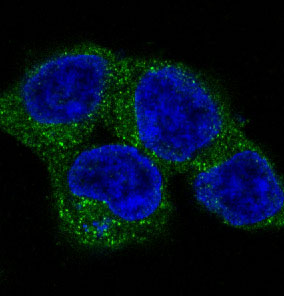
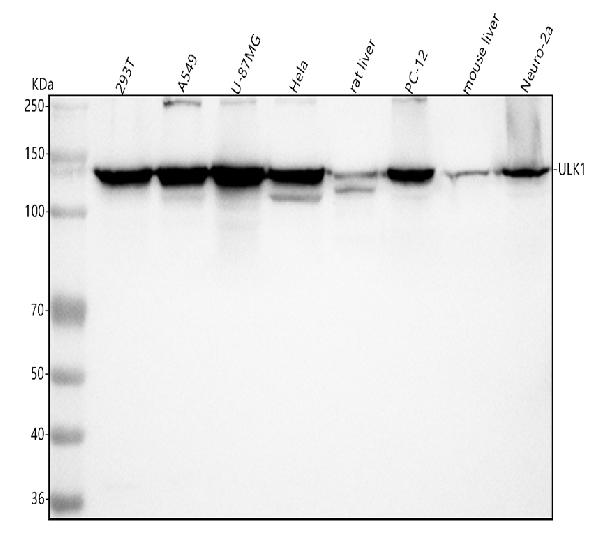
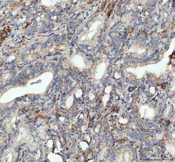
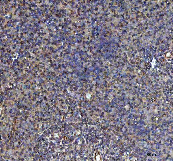
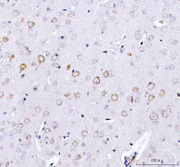
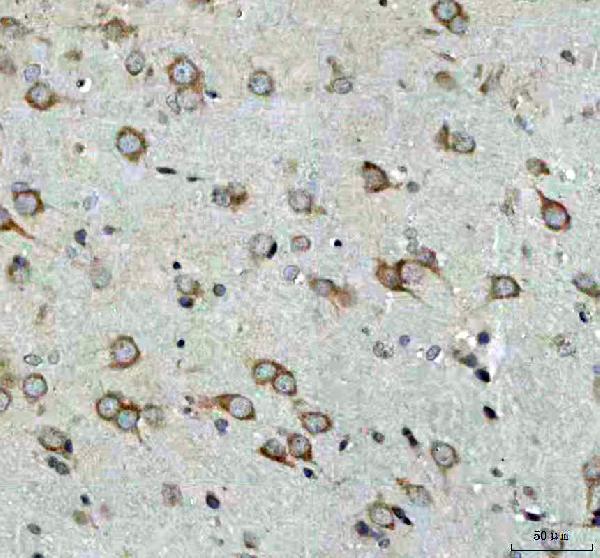
Anti-ULK1/Atg1 Rabbit Monoclonal Antibody
- SPECIFICATION
- CITATIONS
- PROTOCOLS
- BACKGROUND

Application
| WB, IHC, IF, ICC |
|---|---|
| Primary Accession | O75385 |
| Host | Rabbit |
| Isotype | Rabbit IgG |
| Reactivity | Rat, Human, Mouse |
| Clonality | Monoclonal |
| Format | Liquid |
| Description | Anti-ULK1/Atg1 Rabbit Monoclonal Antibody . Tested in WB, IHC, ICC/IF applications. This antibody reacts with Human, Mouse, Rat. |
| Gene ID | 8408 |
|---|---|
| Other Names | Serine/threonine-protein kinase ULK1, 2.7.11.1, Autophagy-related protein 1 homolog, ATG1, hATG1, Unc-51-like kinase 1, ULK1 {ECO:0000303|PubMed:9693035, ECO:0000312|HGNC:HGNC:12558} |
| Calculated MW | 112631 MW KDa |
| Application Details | WB 1:500-1:2000 IHC 1:50-1:200 ICC/IF 1:50-1:200 |
| Subcellular Localization | Cytoplasm, cytosol. Preautophagosomal structure. Under starvation conditions, is localized to puncate structures primarily representing the isolation membrane that sequesters a portion of the cytoplasm resulting in the formation of an autophagosome.. |
| Tissue Specificity | Ubiquitously expressed. Detected in the following adult tissues: skeletal muscle, heart, pancreas, brain, placenta, liver, kidney, and lung. |
| Contents | Rabbit IgG in phosphate buffered saline, pH 7.4, 150mM NaCl, 0.02% sodium azide and 50% glycerol, 0.4-0.5mg/ml BSA. |
| Clone Names | Clone: ABHH-21 |
| Immunogen | A synthesized peptide derived from human ULK1 |
| Purification | Affinity-chromatography |
| Storage | Store at -20°C for one year. For short term storage and frequent use, store at 4°C for up to one month. Avoid repeated freeze-thaw cycles. |
| Name | ULK1 {ECO:0000303|PubMed:9693035, ECO:0000312|HGNC:HGNC:12558} |
|---|---|
| Function | Serine/threonine-protein kinase involved in autophagy in response to starvation (PubMed:18936157, PubMed:21460634, PubMed:21795849, PubMed:23524951, PubMed:25040165, PubMed:29487085, PubMed:31123703). Acts upstream of phosphatidylinositol 3-kinase PIK3C3 to regulate the formation of autophagophores, the precursors of autophagosomes (PubMed:18936157, PubMed:21460634, PubMed:21795849, PubMed:25040165). Part of regulatory feedback loops in autophagy: acts both as a downstream effector and negative regulator of mammalian target of rapamycin complex 1 (mTORC1) via interaction with RPTOR (PubMed:21795849). Activated via phosphorylation by AMPK and also acts as a regulator of AMPK by mediating phosphorylation of AMPK subunits PRKAA1, PRKAB2 and PRKAG1, leading to negatively regulate AMPK activity (PubMed:21460634). May phosphorylate ATG13/KIAA0652 and RPTOR; however such data need additional evidences (PubMed:18936157). Plays a role early in neuronal differentiation and is required for granule cell axon formation (PubMed:11146101). Also phosphorylates SESN2 and SQSTM1 to regulate autophagy (PubMed:25040165, PubMed:37306101). Phosphorylates FLCN, promoting autophagy (PubMed:25126726). Phosphorylates AMBRA1 in response to autophagy induction, releasing AMBRA1 from the cytoskeletal docking site to induce autophagosome nucleation (PubMed:20921139). Phosphorylates ATG4B, leading to inhibit autophagy by decreasing both proteolytic activation and delipidation activities of ATG4B (PubMed:28821708). |
| Cellular Location | Cytoplasm, cytosol. Preautophagosomal structure. Note=Under starvation conditions, is localized to puncate structures primarily representing the isolation membrane that sequesters a portion of the cytoplasm resulting in the formation of an autophagosome. |
| Tissue Location | Ubiquitously expressed. Detected in the following adult tissues: skeletal muscle, heart, pancreas, brain, placenta, liver, kidney, and lung |
Research Areas
Citations (0)

Thousands of laboratories across the world have published research that depended on the performance of antibodies from Abcepta to advance their research. Check out links to articles that cite our products in major peer-reviewed journals, organized by research category.
Submit your citation using an Abcepta antibody to
info@abcepta.com, and receive a free "I Love Antibodies" mug.
info@abcepta.com, and receive a free "I Love Antibodies" mug.

Application Protocols
Provided below are standard protocols that you may find useful for product applications.
Abcepta welcomes feedback from its customers.
If you have used an Abcepta product and would like to share how it has performed, please click on the "Submit Review" button and provide the requested information. Our staff will examine and post your review and contact you if needed.
If you have any additional inquiries please email technical services at tech@abcepta.com.
$ 370.00
Cat# ABO13565
Ordering Information
United States
AlbaniaAustraliaAustriaBelgiumBosnia & HerzegovinaBrazilBulgariaCanadaCentral AmericaChinaCroatiaCyprusCzech RepublicDenmarkEstoniaFinlandFranceGermanyGreeceHong KongHungaryIcelandIndiaIndonesiaIrelandIsraelItalyJapanLatviaLithuaniaLuxembourgMacedoniaMalaysiaMaltaNetherlandsNew ZealandNorwayPakistanPolandPortugalRomaniaSerbiaSingaporeSlovakiaSloveniaSouth AfricaSouth KoreaSpainSwedenSwitzerlandTaiwanTurkeyUnited KingdomUnited StatesVietnamWorldwideOthersMexico
USA Headquarters
(888) 735-7227 / (858) 622-0099 or (858) 875-1900
Other Products
Shipping Information
Domestic orders (in stock items)
Shipped out the same day. Orders placed after 1 PM (PST) will ship out the next business day.
International orders
Contact your local distributors
