


 Foundational characteristics of cancer include proliferation, angiogenesis, migration, evasion of apoptosis, and cellular immortality. Find key markers for these cellular processes and antibodies to detect them.
 The SUMOplot™ Analysis Program predicts and scores sumoylation sites in your protein. SUMOylation is a post-translational modification involved in various cellular processes, such as nuclear-cytosolic transport, transcriptional regulation, apoptosis, protein stability, response to stress, and progression through the cell cycle.
 The Autophagy Receptor Motif Plotter predicts and scores autophagy receptor binding sites in your protein. Identifying proteins connected to this pathway is critical to understanding the role of autophagy in physiological as well as pathological processes such as development, differentiation, neurodegenerative diseases, stress, infection, and cancer.
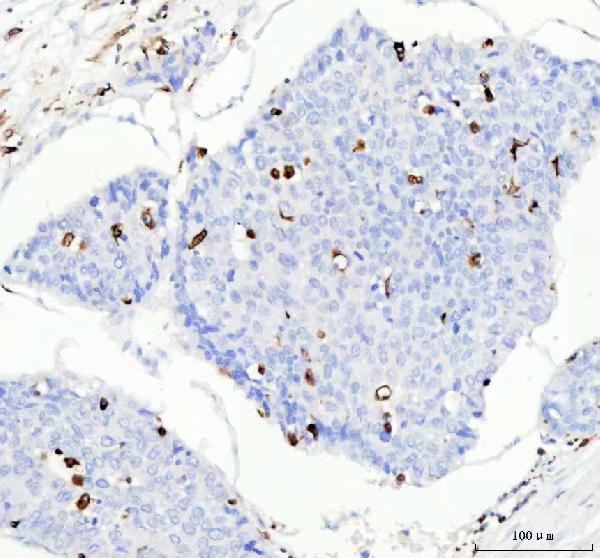
Anti-CD74 Rabbit Monoclonal Antibody

- SPECIFICATION
- CITATIONS
- PROTOCOLS
- BACKGROUND

Application
| WB, IHC, IF, ICC, FC |
|---|---|
| Primary Accession | P04233 |
| Host | Rabbit |
| Isotype | Rabbit IgG |
| Reactivity | Human |
| Clonality | Monoclonal |
| Format | Liquid |
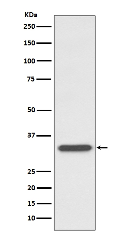
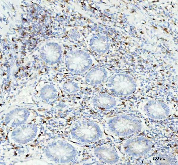
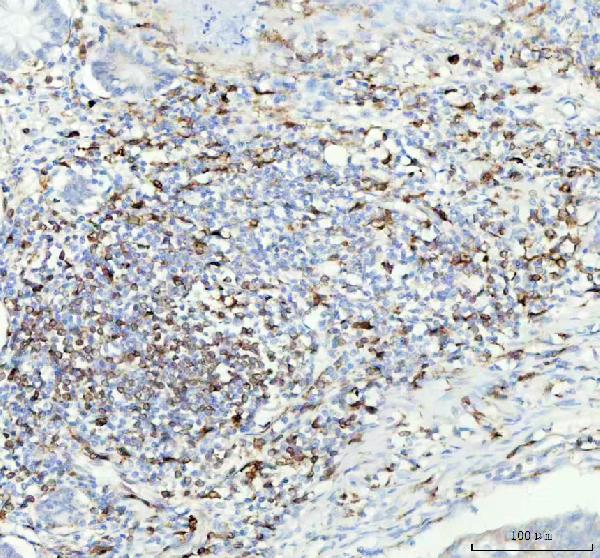
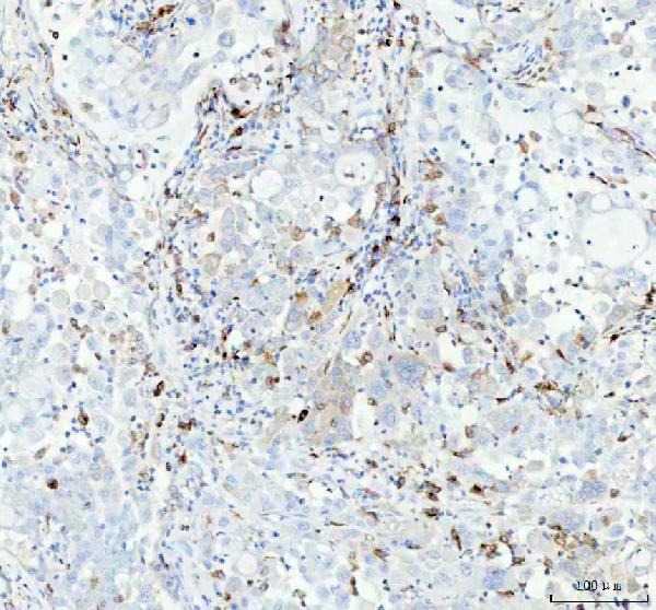
| Description | Anti-CD74 Rabbit Monoclonal Antibody . Tested in WB, IHC, ICC/IF, Flow Cytometry applications. This antibody reacts with Human. |
| Gene ID | 972 |
|---|---|
| Other Names | HLA class II histocompatibility antigen gamma chain, HLA-DR antigens-associated invariant chain, Ia antigen-associated invariant chain, Ii, CD74, Class-II-associated invariant chain peptide, CLIP, CD74 (HGNC:1697), DHLAG |
| Calculated MW | 33516 MW KDa |
| Application Details | WB 1:500-1:2000 IHC 1:50-1:200 ICC/IF 1:50-1:200 FC 1:50 |
| Subcellular Localization | Cell membrane ; Single-pass type II membrane protein. Endoplasmic reticulum membrane. Golgi apparatus, trans-Golgi network. Endosome. Lysosome. Transits through a number of intracellular compartments in the endocytic pathway. It can either undergo proteolysis or reach the cell membrane. |
| Contents | Rabbit IgG in phosphate buffered saline, pH 7.4, 150mM NaCl, 0.02% sodium azide and 50% glycerol, 0.4-0.5mg/ml BSA. |
| Clone Names | Clone: AOIF-3 |
| Immunogen | A synthesized peptide derived from human CD74 |
| Purification | Affinity-chromatography |
| Storage | Store at -20°C for one year. For short term storage and frequent use, store at 4°C for up to one month. Avoid repeated freeze-thaw cycles. |
| Name | CD74 (HGNC:1697) |
|---|---|
| Synonyms | DHLAG |
| Function | Plays a critical role in MHC class II antigen processing by stabilizing peptide-free class II alpha/beta heterodimers in a complex soon after their synthesis and directing transport of the complex from the endoplasmic reticulum to the endosomal/lysosomal system where the antigen processing and binding of antigenic peptides to MHC class II takes place. Serves as cell surface receptor for the cytokine MIF. [Isoform p41]: Stabilizes the conformation of mature CTSL by binding to its active site and serving as a chaperone to help maintain a pool of mature enzyme in endocytic compartments and extracellular space of antigen-presenting cells (APCs). Has antiviral activity by stymieing the endosomal entry of Ebola virus and coronaviruses, including SARS-CoV-2 (PubMed:32855215). Disrupts cathepsin-mediated Ebola virus glycoprotein processing, which prevents viral fusion and entry. This antiviral activity is specific to p41 isoform (PubMed:32855215). |
| Cellular Location | Cell membrane; Single-pass type II membrane protein. Endoplasmic reticulum membrane. Golgi apparatus, trans-Golgi network. Endosome. Lysosome. Secreted. Note=Transits through a number of intracellular compartments in the endocytic pathway. It can either undergo proteolysis or reach the cell membrane |
| Tissue Location | Detected in urine (at protein level). [Isoform p33]: In B cells, represents 70% of total CD74 expression. |
Research Areas
Citations (0)

Thousands of laboratories across the world have published research that depended on the performance of antibodies from Abcepta to advance their research. Check out links to articles that cite our products in major peer-reviewed journals, organized by research category.
Submit your citation using an Abcepta antibody to
info@abcepta.com, and receive a free "I Love Antibodies" mug.
info@abcepta.com, and receive a free "I Love Antibodies" mug.

Application Protocols
Provided below are standard protocols that you may find useful for product applications.
Abcepta welcomes feedback from its customers.
If you have used an Abcepta product and would like to share how it has performed, please click on the "Submit Review" button and provide the requested information. Our staff will examine and post your review and contact you if needed.
If you have any additional inquiries please email technical services at tech@abcepta.com.
$ 370.00
Cat# ABO13520
Ordering Information
United States
AlbaniaAustraliaAustriaBelgiumBosnia & HerzegovinaBrazilBulgariaCanadaCentral AmericaChinaCroatiaCyprusCzech RepublicDenmarkEstoniaFinlandFranceGermanyGreeceHong KongHungaryIcelandIndiaIndonesiaIrelandIsraelItalyJapanLatviaLithuaniaLuxembourgMacedoniaMalaysiaMaltaNetherlandsNew ZealandNorwayPakistanPolandPortugalRomaniaSerbiaSingaporeSlovakiaSloveniaSouth AfricaSouth KoreaSpainSwedenSwitzerlandTaiwanTurkeyUnited KingdomUnited StatesVietnamWorldwideOthersMexico
USA Headquarters
(888) 735-7227 / (858) 622-0099 or (858) 875-1900
Other Products
Shipping Information
Domestic orders (in stock items)
Shipped out the same day. Orders placed after 1 PM (PST) will ship out the next business day.
International orders
Contact your local distributors
