


 Foundational characteristics of cancer include proliferation, angiogenesis, migration, evasion of apoptosis, and cellular immortality. Find key markers for these cellular processes and antibodies to detect them.
 The SUMOplot™ Analysis Program predicts and scores sumoylation sites in your protein. SUMOylation is a post-translational modification involved in various cellular processes, such as nuclear-cytosolic transport, transcriptional regulation, apoptosis, protein stability, response to stress, and progression through the cell cycle.
 The Autophagy Receptor Motif Plotter predicts and scores autophagy receptor binding sites in your protein. Identifying proteins connected to this pathway is critical to understanding the role of autophagy in physiological as well as pathological processes such as development, differentiation, neurodegenerative diseases, stress, infection, and cancer.
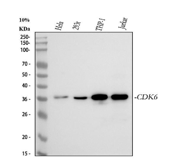
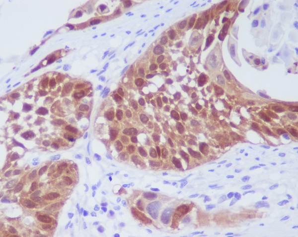
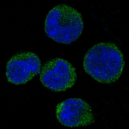
Anti-CDK6 Rabbit Monoclonal Antibody
- SPECIFICATION
- CITATIONS
- PROTOCOLS
- BACKGROUND

Application
| WB, IHC, IF, ICC, FC |
|---|---|
| Primary Accession | Q00534 |
| Host | Rabbit |
| Isotype | Rabbit IgG |
| Reactivity | Human |
| Clonality | Monoclonal |
| Format | Liquid |
| Description | Anti-CDK6 Rabbit Monoclonal Antibody . Tested in WB, IHC, ICC/IF, Flow Cytometry applications. This antibody reacts with Human. |
| Gene ID | 1021 |
|---|---|
| Other Names | Cyclin-dependent kinase 6, 2.7.11.22, Cell division protein kinase 6, Serine/threonine-protein kinase PLSTIRE, CDK6, CDKN6 |
| Calculated MW | 36938 MW KDa |
| Application Details | WB 1:5000-1:20000 IHC 1:50-1:200 ICC/IF 1:50-1:200 FC 1:100 |
| Subcellular Localization | Cytoplasm. Nucleus. Cell projection, ruffle. Cytoplasm, cytoskeleton, microtubule organizing center, centrosome. Localized to the ruffling edge of spreading fibroblasts. Kinase activity only in nucleus. Localized to the cytosol of neurons and showed prominent staining around either side of the nucleus (By similarity). Present in the cytosol and in the nucleus in interphase cells and at the centrosome during mitosis from prophase to telophase (PubMed:23918663).. |
| Tissue Specificity | Expressed ubiquitously. Accumulates in squamous cell carcinomas, proliferating hematopoietic progenitor cells, beta-cells of pancreatic islets of Langerhans, and neuroblastomas. Reduced levels in differentiating cells.. |
| Contents | Rabbit IgG in phosphate buffered saline, pH 7.4, 150mM NaCl, 0.02% sodium azide and 50% glycerol, 0.4-0.5mg/ml BSA. |
| Clone Names | Clone: HOA-3 |
| Immunogen | A synthesized peptide derived from human CDK6 |
| Purification | Affinity-chromatography |
| Storage | Store at -20°C for one year. For short term storage and frequent use, store at 4°C for up to one month. Avoid repeated freeze-thaw cycles. |
| Name | CDK6 |
|---|---|
| Synonyms | CDKN6 |
| Function | Serine/threonine-protein kinase involved in the control of the cell cycle and differentiation; promotes G1/S transition. Phosphorylates pRB/RB1 and NPM1. Interacts with D-type G1 cyclins during interphase at G1 to form a pRB/RB1 kinase and controls the entrance into the cell cycle. Involved in initiation and maintenance of cell cycle exit during cell differentiation; prevents cell proliferation and negatively regulates cell differentiation, but is required for the proliferation of specific cell types (e.g. erythroid and hematopoietic cells). Essential for cell proliferation within the dentate gyrus of the hippocampus and the subventricular zone of the lateral ventricles. Required during thymocyte development. Promotes the production of newborn neurons, probably by modulating G1 length. Promotes, at least in astrocytes, changes in patterns of gene expression, changes in the actin cytoskeleton including loss of stress fibers, and enhanced motility during cell differentiation. Prevents myeloid differentiation by interfering with RUNX1 and reducing its transcription transactivation activity, but promotes proliferation of normal myeloid progenitors. Delays senescence. Promotes the proliferation of beta-cells in pancreatic islets of Langerhans. May play a role in the centrosome organization during the cell cycle phases (PubMed:23918663). |
| Cellular Location | Cytoplasm. Nucleus. Cell projection, ruffle. Cytoplasm, cytoskeleton, microtubule organizing center, centrosome. Note=Localized to the ruffling edge of spreading fibroblasts. Kinase activity only in nucleus. Localized to the cytosol of neurons and showed prominent staining around either side of the nucleus (By similarity). Present in the cytosol and in the nucleus in interphase cells and at the centrosome during mitosis from prophase to telophase (PubMed:23918663). {ECO:0000250|UniProtKB:Q64261, ECO:0000269|PubMed:23918663} |
| Tissue Location | Expressed ubiquitously. Accumulates in squamous cell carcinomas, proliferating hematopoietic progenitor cells, beta- cells of pancreatic islets of Langerhans, and neuroblastomas. Reduced levels in differentiating cells. |
Research Areas
Citations (0)

Thousands of laboratories across the world have published research that depended on the performance of antibodies from Abcepta to advance their research. Check out links to articles that cite our products in major peer-reviewed journals, organized by research category.
Submit your citation using an Abcepta antibody to
info@abcepta.com, and receive a free "I Love Antibodies" mug.
info@abcepta.com, and receive a free "I Love Antibodies" mug.

Application Protocols
Provided below are standard protocols that you may find useful for product applications.
Abcepta welcomes feedback from its customers.
If you have used an Abcepta product and would like to share how it has performed, please click on the "Submit Review" button and provide the requested information. Our staff will examine and post your review and contact you if needed.
If you have any additional inquiries please email technical services at tech@abcepta.com.
$ 370.00
Cat# ABO13457
Ordering Information
United States
AlbaniaAustraliaAustriaBelgiumBosnia & HerzegovinaBrazilBulgariaCanadaCentral AmericaChinaCroatiaCyprusCzech RepublicDenmarkEstoniaFinlandFranceGermanyGreeceHong KongHungaryIcelandIndiaIndonesiaIrelandIsraelItalyJapanLatviaLithuaniaLuxembourgMacedoniaMalaysiaMaltaNetherlandsNew ZealandNorwayPakistanPolandPortugalRomaniaSerbiaSingaporeSlovakiaSloveniaSouth AfricaSouth KoreaSpainSwedenSwitzerlandTaiwanTurkeyUnited KingdomUnited StatesVietnamWorldwideOthersMexico
USA Headquarters
(888) 735-7227 / (858) 622-0099 or (858) 875-1900
Other Products
Shipping Information
Domestic orders (in stock items)
Shipped out the same day. Orders placed after 1 PM (PST) will ship out the next business day.
International orders
Contact your local distributors
