


 Foundational characteristics of cancer include proliferation, angiogenesis, migration, evasion of apoptosis, and cellular immortality. Find key markers for these cellular processes and antibodies to detect them.
 The SUMOplot™ Analysis Program predicts and scores sumoylation sites in your protein. SUMOylation is a post-translational modification involved in various cellular processes, such as nuclear-cytosolic transport, transcriptional regulation, apoptosis, protein stability, response to stress, and progression through the cell cycle.
 The Autophagy Receptor Motif Plotter predicts and scores autophagy receptor binding sites in your protein. Identifying proteins connected to this pathway is critical to understanding the role of autophagy in physiological as well as pathological processes such as development, differentiation, neurodegenerative diseases, stress, infection, and cancer.
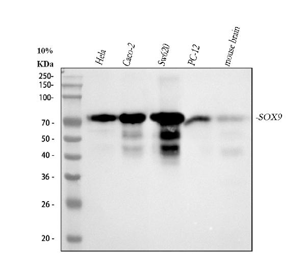
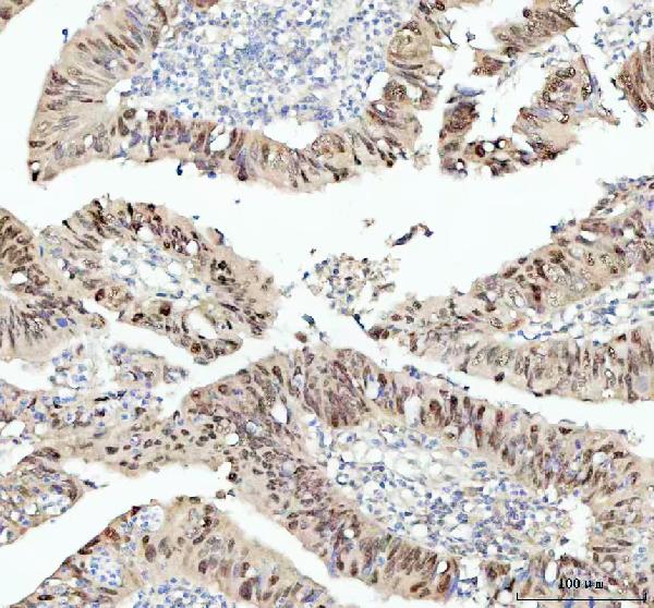
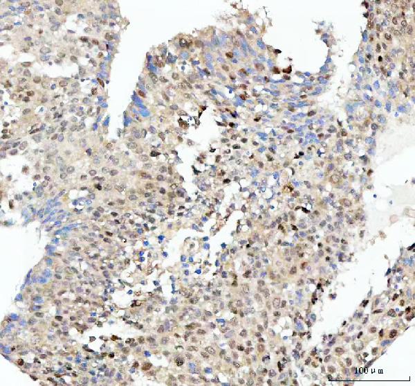
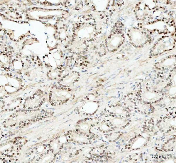
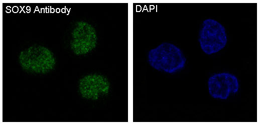
Anti-SOX9 Rabbit Monoclonal Antibody
- SPECIFICATION
- CITATIONS
- PROTOCOLS
- BACKGROUND

Application
| WB, IHC, IF, ICC |
|---|---|
| Primary Accession | P48436 |
| Host | Rabbit |
| Isotype | Rabbit IgG |
| Reactivity | Rat, Human, Mouse |
| Clonality | Monoclonal |
| Format | Liquid |
| Description | Anti-SOX9 Rabbit Monoclonal Antibody . Tested in WB, IHC, ICC/IF applications. This antibody reacts with Human, Mouse, Rat. |
| Gene ID | 6662 |
|---|---|
| Other Names | Transcription factor SOX-9, SOX9 {ECO:0000303|PubMed:7990924, ECO:0000312|HGNC:HGNC:11204} |
| Calculated MW | 56137 MW KDa |
| Application Details | WB 1:500-1:2000 IHC 1:100-1:500 ICC/IF 1:50-1:200 |
| Subcellular Localization | Nucleus. |
| Contents | Rabbit IgG in phosphate buffered saline, pH 7.4, 150mM NaCl, 0.02% sodium azide and 50% glycerol, 0.4-0.5mg/ml BSA. |
| Clone Names | Clone: DAO-19 |
| Immunogen | A synthesized peptide derived from human SOX9 |
| Purification | Affinity-chromatography |
| Storage | Store at -20°C for one year. For short term storage and frequent use, store at 4°C for up to one month. Avoid repeated freeze-thaw cycles. |
| Name | SOX9 {ECO:0000303|PubMed:7990924, ECO:0000312|HGNC:HGNC:11204} |
|---|---|
| Function | Transcription factor that plays a key role in chondrocytes differentiation and skeletal development (PubMed:24038782). Specifically binds the 5'-ACAAAG-3' DNA motif present in enhancers and super-enhancers and promotes expression of genes important for chondrogenesis, including cartilage matrix protein-coding genes COL2A1, COL4A2, COL9A1, COL11A2 and ACAN, SOX5 and SOX6 (PubMed:8640233). Also binds to some promoter regions (By similarity). Plays a central role in successive steps of chondrocyte differentiation (By similarity). Absolutely required for precartilaginous condensation, the first step in chondrogenesis during which skeletal progenitors differentiate into prechondrocytes (By similarity). Together with SOX5 and SOX6, required for overt chondrogenesis when condensed prechondrocytes differentiate into early stage chondrocytes, the second step in chondrogenesis (By similarity). Later, required to direct hypertrophic maturation and block osteoblast differentiation of growth plate chondrocytes: maintains chondrocyte columnar proliferation, delays prehypertrophy and then prevents osteoblastic differentiation of chondrocytes by lowering beta-catenin (CTNNB1) signaling and RUNX2 expression (By similarity). Also required for chondrocyte hypertrophy, both indirectly, by keeping the lineage fate of chondrocytes, and directly, by remaining present in upper hypertrophic cells and transactivating COL10A1 along with MEF2C (By similarity). Low lipid levels are the main nutritional determinant for chondrogenic commitment of skeletal progenitor cells: when lipids levels are low, FOXO (FOXO1 and FOXO3) transcription factors promote expression of SOX9, which induces chondrogenic commitment and suppresses fatty acid oxidation (By similarity). Mechanistically, helps, but is not required, to remove epigenetic signatures of transcriptional repression and deposit active promoter and enhancer marks at chondrocyte-specific genes (By similarity). Acts in cooperation with the Hedgehog pathway-dependent GLI (GLI1 and GLI3) transcription factors (By similarity). In addition to cartilage development, also acts as a regulator of proliferation and differentiation in epithelial stem/progenitor cells: involved in the lung epithelium during branching morphogenesis, by balancing proliferation and differentiation and regulating the extracellular matrix (By similarity). Controls epithelial branching during kidney development (By similarity). |
| Cellular Location | Nucleus {ECO:0000255|PROSITE-ProRule:PRU00267, ECO:0000269|PubMed:8640233} |
Research Areas
Citations (0)

Thousands of laboratories across the world have published research that depended on the performance of antibodies from Abcepta to advance their research. Check out links to articles that cite our products in major peer-reviewed journals, organized by research category.
Submit your citation using an Abcepta antibody to
info@abcepta.com, and receive a free "I Love Antibodies" mug.
info@abcepta.com, and receive a free "I Love Antibodies" mug.

Application Protocols
Provided below are standard protocols that you may find useful for product applications.
Abcepta welcomes feedback from its customers.
If you have used an Abcepta product and would like to share how it has performed, please click on the "Submit Review" button and provide the requested information. Our staff will examine and post your review and contact you if needed.
If you have any additional inquiries please email technical services at tech@abcepta.com.
$ 370.00
Cat# ABO13366
Ordering Information
United States
AlbaniaAustraliaAustriaBelgiumBosnia & HerzegovinaBrazilBulgariaCanadaCentral AmericaChinaCroatiaCyprusCzech RepublicDenmarkEstoniaFinlandFranceGermanyGreeceHong KongHungaryIcelandIndiaIndonesiaIrelandIsraelItalyJapanLatviaLithuaniaLuxembourgMacedoniaMalaysiaMaltaNetherlandsNew ZealandNorwayPakistanPolandPortugalRomaniaSerbiaSingaporeSlovakiaSloveniaSouth AfricaSouth KoreaSpainSwedenSwitzerlandTaiwanTurkeyUnited KingdomUnited StatesVietnamWorldwideOthersMexico
USA Headquarters
(888) 735-7227 / (858) 622-0099 or (858) 875-1900
Other Products
Shipping Information
Domestic orders (in stock items)
Shipped out the same day. Orders placed after 1 PM (PST) will ship out the next business day.
International orders
Contact your local distributors
