


 Foundational characteristics of cancer include proliferation, angiogenesis, migration, evasion of apoptosis, and cellular immortality. Find key markers for these cellular processes and antibodies to detect them.
 The SUMOplot™ Analysis Program predicts and scores sumoylation sites in your protein. SUMOylation is a post-translational modification involved in various cellular processes, such as nuclear-cytosolic transport, transcriptional regulation, apoptosis, protein stability, response to stress, and progression through the cell cycle.
 The Autophagy Receptor Motif Plotter predicts and scores autophagy receptor binding sites in your protein. Identifying proteins connected to this pathway is critical to understanding the role of autophagy in physiological as well as pathological processes such as development, differentiation, neurodegenerative diseases, stress, infection, and cancer.
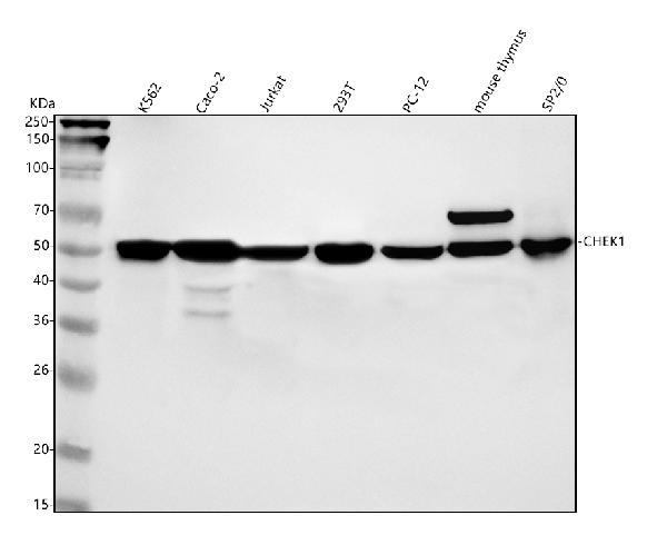
Anti-Chk1 CHEK1 Rabbit Monoclonal Antibody
- SPECIFICATION
- CITATIONS
- PROTOCOLS
- BACKGROUND

Application
| WB, IF, ICC, IP |
|---|---|
| Primary Accession | O14757 |
| Host | Rabbit |
| Isotype | Rabbit IgG |
| Reactivity | Rat, Human, Mouse |
| Clonality | Monoclonal |
| Format | Liquid |
| Description | Anti-Chk1 CHEK1 Rabbit Monoclonal Antibody . Tested in WB, ICC/IF, IP applications. This antibody reacts with Human, Mouse, Rat. |
| Gene ID | 1111 |
|---|---|
| Other Names | Serine/threonine-protein kinase Chk1, 2.7.11.1, CHK1 checkpoint homolog, Cell cycle checkpoint kinase, Checkpoint kinase-1, CHEK1, CHK1 |
| Calculated MW | 54434 MW KDa |
| Application Details | WB 1:500-1:2000 ICC/IF 1:50-1:200 IP 1:30 |
| Subcellular Localization | Nucleus. Cytoplasm. Cytoplasm, cytoskeleton, microtubule organizing center, centrosome. Nuclear export is mediated at least in part by XPO1/CRM1. Also localizes to the centrosome specifically during interphase, where it may protect centrosomal CDC2 kinase from inappropriate activation by cytoplasmic CDC25B. |
| Tissue Specificity | Expressed ubiquitously with the most abundant expression in thymus, testis, small intestine and colon.. |
| Contents | Rabbit IgG in phosphate buffered saline, pH 7.4, 150mM NaCl, 0.02% sodium azide and 50% glycerol, 0.4-0.5mg/ml BSA. |
| Clone Names | Clone: AAF-3 |
| Immunogen | A synthesized peptide derived from human Chk1 |
| Purification | Affinity-chromatography |
| Storage | Store at -20°C for one year. For short term storage and frequent use, store at 4°C for up to one month. Avoid repeated freeze-thaw cycles. |
| Name | CHEK1 |
|---|---|
| Synonyms | CHK1 |
| Function | Serine/threonine-protein kinase which is required for checkpoint-mediated cell cycle arrest and activation of DNA repair in response to the presence of DNA damage or unreplicated DNA (PubMed:11535615, PubMed:12399544, PubMed:12446774, PubMed:14559997, PubMed:14988723, PubMed:15311285, PubMed:15650047, PubMed:15665856, PubMed:32357935). May also negatively regulate cell cycle progression during unperturbed cell cycles (PubMed:11535615, PubMed:12399544, PubMed:12446774, PubMed:14559997, PubMed:14988723, PubMed:15311285, PubMed:15650047, PubMed:15665856). This regulation is achieved by a number of mechanisms that together help to preserve the integrity of the genome (PubMed:11535615, PubMed:12399544, PubMed:12446774, PubMed:14559997, PubMed:14988723, PubMed:15311285, PubMed:15650047, PubMed:15665856). Recognizes the substrate consensus sequence [R-X-X- S/T] (PubMed:11535615, PubMed:12399544, PubMed:12446774, PubMed:14559997, PubMed:14988723, PubMed:15311285, PubMed:15650047, PubMed:15665856). Binds to and phosphorylates CDC25A, CDC25B and CDC25C (PubMed:12676583, PubMed:12676925, PubMed:12759351, PubMed:14559997, PubMed:14681206, PubMed:19734889, PubMed:9278511). Phosphorylation of CDC25A at 'Ser-178' and 'Thr-507' and phosphorylation of CDC25C at 'Ser-216' creates binding sites for 14-3-3 proteins which inhibit CDC25A and CDC25C (PubMed:9278511). Phosphorylation of CDC25A at 'Ser- 76', 'Ser-124', 'Ser-178', 'Ser-279' and 'Ser-293' promotes proteolysis of CDC25A (PubMed:12676583, PubMed:12676925, PubMed:12759351, PubMed:14681206, PubMed:19734889, PubMed:9278511). Phosphorylation of CDC25A at 'Ser-76' primes the protein for subsequent phosphorylation at 'Ser-79', 'Ser-82' and 'Ser-88' by NEK11, which is required for polyubiquitination and degradation of CDCD25A (PubMed:19734889, PubMed:20090422, PubMed:9278511). Inhibition of CDC25 leads to increased inhibitory tyrosine phosphorylation of CDK-cyclin complexes and blocks cell cycle progression (PubMed:9278511). Also phosphorylates NEK6 (PubMed:18728393). Binds to and phosphorylates RAD51 at 'Thr-309', which promotes the release of RAD51 from BRCA2 and enhances the association of RAD51 with chromatin, thereby promoting DNA repair by homologous recombination (PubMed:15665856). Phosphorylates multiple sites within the C-terminus of TP53, which promotes activation of TP53 by acetylation and promotes cell cycle arrest and suppression of cellular proliferation (PubMed:10673501, PubMed:15659650, PubMed:16511572). Also promotes repair of DNA cross-links through phosphorylation of FANCE (PubMed:17296736). Binds to and phosphorylates TLK1 at 'Ser-743', which prevents the TLK1-dependent phosphorylation of the chromatin assembly factor ASF1A (PubMed:12660173, PubMed:12955071). This may enhance chromatin assembly both in the presence or absence of DNA damage (PubMed:12660173, PubMed:12955071). May also play a role in replication fork maintenance through regulation of PCNA (PubMed:18451105). May regulate the transcription of genes that regulate cell-cycle progression through the phosphorylation of histones (By similarity). Phosphorylates histone H3.1 (to form H3T11ph), which leads to epigenetic inhibition of a subset of genes (By similarity). May also phosphorylate RB1 to promote its interaction with the E2F family of transcription factors and subsequent cell cycle arrest (PubMed:17380128). Phosphorylates SPRTN, promoting SPRTN recruitment to chromatin (PubMed:31316063). Reduces replication stress and activates the G2/M checkpoint, by phosphorylating and inactivating PABIR1/FAM122A and promoting the serine/threonine-protein phosphatase 2A-mediated dephosphorylation and stabilization of WEE1 levels and activity (PubMed:33108758). |
| Cellular Location | Nucleus. Chromosome. Cytoplasm Cytoplasm, cytoskeleton, microtubule organizing center, centrosome. Note=Nuclear export is mediated at least in part by XPO1/CRM1 (PubMed:12676962). Also localizes to the centrosome specifically during interphase, where it may protect centrosomal CDC2 kinase from inappropriate activation by cytoplasmic CDC25B (PubMed:15311285). Proteolytic cleavage at the C-terminus by SPRTN promotes removal from chromatin (PubMed:31316063) |
| Tissue Location | Expressed ubiquitously with the most abundant expression in thymus, testis, small intestine and colon |
Research Areas
Citations (0)

Thousands of laboratories across the world have published research that depended on the performance of antibodies from Abcepta to advance their research. Check out links to articles that cite our products in major peer-reviewed journals, organized by research category.
Submit your citation using an Abcepta antibody to
info@abcepta.com, and receive a free "I Love Antibodies" mug.
info@abcepta.com, and receive a free "I Love Antibodies" mug.

Application Protocols
Provided below are standard protocols that you may find useful for product applications.
Abcepta welcomes feedback from its customers.
If you have used an Abcepta product and would like to share how it has performed, please click on the "Submit Review" button and provide the requested information. Our staff will examine and post your review and contact you if needed.
If you have any additional inquiries please email technical services at tech@abcepta.com.
$ 370.00
Cat# ABO13299
Ordering Information
United States
AlbaniaAustraliaAustriaBelgiumBosnia & HerzegovinaBrazilBulgariaCanadaCentral AmericaChinaCroatiaCyprusCzech RepublicDenmarkEstoniaFinlandFranceGermanyGreeceHong KongHungaryIcelandIndiaIndonesiaIrelandIsraelItalyJapanLatviaLithuaniaLuxembourgMacedoniaMalaysiaMaltaNetherlandsNew ZealandNorwayPakistanPolandPortugalRomaniaSerbiaSingaporeSlovakiaSloveniaSouth AfricaSouth KoreaSpainSwedenSwitzerlandTaiwanTurkeyUnited KingdomUnited StatesVietnamWorldwideOthersMexico
USA Headquarters
(888) 735-7227 / (858) 622-0099 or (858) 875-1900
Other Products
Shipping Information
Domestic orders (in stock items)
Shipped out the same day. Orders placed after 1 PM (PST) will ship out the next business day.
International orders
Contact your local distributors
