


 Foundational characteristics of cancer include proliferation, angiogenesis, migration, evasion of apoptosis, and cellular immortality. Find key markers for these cellular processes and antibodies to detect them.
 The SUMOplot™ Analysis Program predicts and scores sumoylation sites in your protein. SUMOylation is a post-translational modification involved in various cellular processes, such as nuclear-cytosolic transport, transcriptional regulation, apoptosis, protein stability, response to stress, and progression through the cell cycle.
 The Autophagy Receptor Motif Plotter predicts and scores autophagy receptor binding sites in your protein. Identifying proteins connected to this pathway is critical to understanding the role of autophagy in physiological as well as pathological processes such as development, differentiation, neurodegenerative diseases, stress, infection, and cancer.
Anti-CD9 Rabbit Monoclonal Antibody
- SPECIFICATION
- CITATIONS
- PROTOCOLS
- BACKGROUND

Application
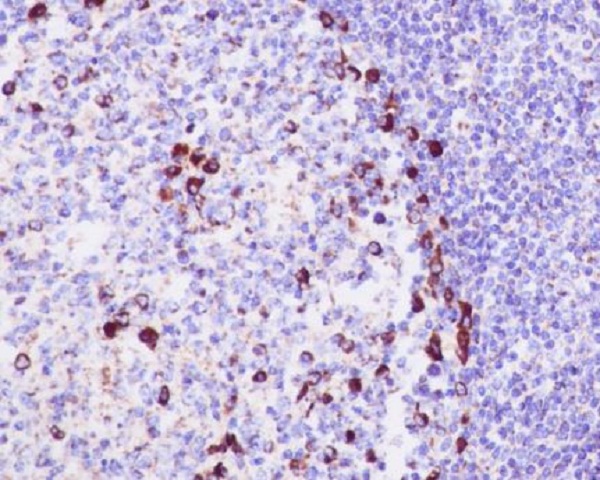
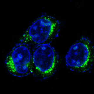
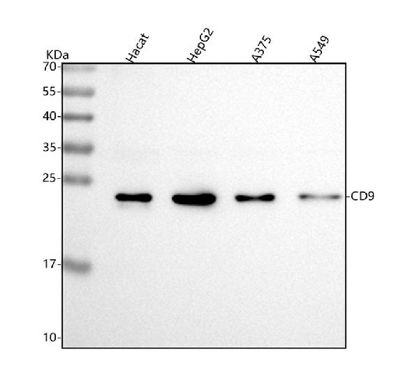
| WB, IHC, IF, ICC, IP, FC |
|---|---|
| Primary Accession | P21926 |
| Host | Rabbit |
| Isotype | Rabbit IgG |
| Reactivity | Rat, Human, Mouse |
| Clonality | Monoclonal |
| Format | Liquid |
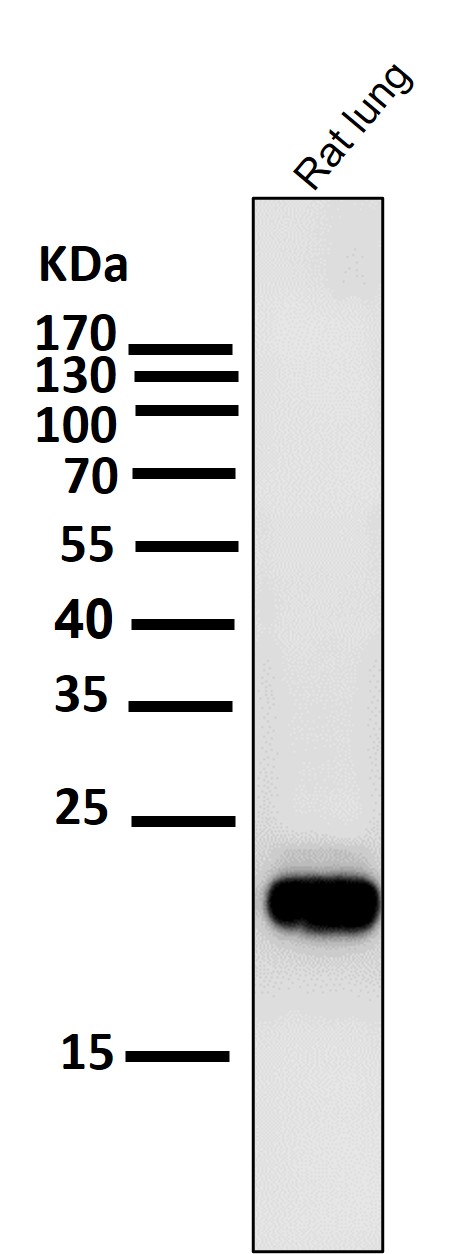
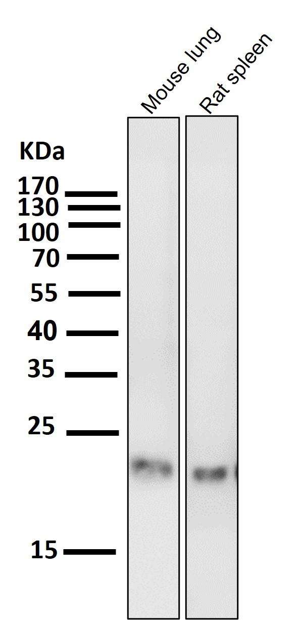
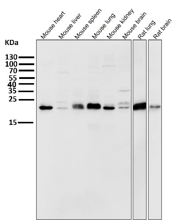
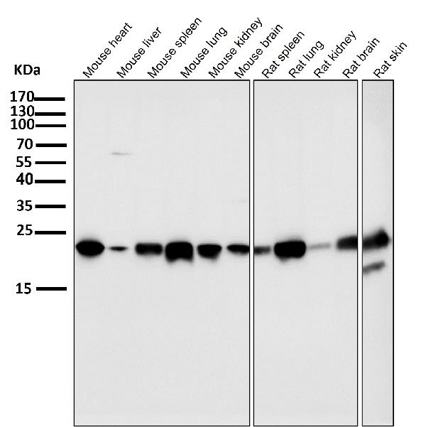
| Description | Anti-CD9 Rabbit Monoclonal Antibody . Tested in WB, IHC, ICC/IF, IP, Flow Cytometry applications. This antibody reacts with Human, Mouse, Rat. |
| Gene ID | 928 |
|---|---|
| Other Names | CD9 antigen, 5H9 antigen, Cell growth-inhibiting gene 2 protein {ECO:0000303|Ref.6}, Leukocyte antigen MIC3, Motility-related protein, MRP-1, Tetraspanin-29, Tspan-29, p24, CD9, CD9 {ECO:0000303|PubMed:1840589, ECO:0000312|HGNC:HGNC:1709} |
| Calculated MW | 25416 MW KDa |
| Application Details | WB 1:1000-1:5000 IHC 1:100-1:500 ICC/IF 1:50-1:200 IP 1:50 FC 1:50 |
| Subcellular Localization | Membrane ; Multi-pass membrane protein. Cell membrane ; Multi-pass membrane protein. |
| Tissue Specificity | Detected in platelets (at protein level). Expressed by a variety of hematopoietic and epithelial cells.. |
| Contents | Rabbit IgG in phosphate buffered saline, pH 7.4, 150mM NaCl, 0.02% sodium azide and 50% glycerol, 0.4-0.5mg/ml BSA. |
| Clone Names | Clone: CED-3 |
| Immunogen | A synthesized peptide derived from human CD9 |
| Purification | Affinity-chromatography |
| Storage | Store at -20°C for one year. For short term storage and frequent use, store at 4°C for up to one month. Avoid repeated freeze-thaw cycles. |
| Name | CD9 {ECO:0000303|PubMed:1840589, ECO:0000312|HGNC:HGNC:1709} |
|---|---|
| Function | Integral membrane protein associated with integrins, which regulates different processes, such as sperm-egg fusion, platelet activation and aggregation, and cell adhesion (PubMed:14575715, PubMed:18541721, PubMed:8478605). Present at the cell surface of oocytes and plays a key role in sperm-egg fusion, possibly by organizing multiprotein complexes and the morphology of the membrane required for the fusion (By similarity). In myoblasts, associates with CD81 and PTGFRN and inhibits myotube fusion during muscle regeneration (By similarity). In macrophages, associates with CD81 and beta-1 and beta-2 integrins, and prevents macrophage fusion into multinucleated giant cells specialized in ingesting complement-opsonized large particles (PubMed:12796480). Also prevents the fusion between mononuclear cell progenitors into osteoclasts in charge of bone resorption (By similarity). Acts as a receptor for PSG17 (By similarity). Involved in platelet activation and aggregation (PubMed:18541721). Regulates paranodal junction formation (By similarity). Involved in cell adhesion, cell motility and tumor metastasis (PubMed:7511626, PubMed:8478605). |
| Cellular Location | Cell membrane; Multi-pass membrane protein. Membrane; Multi-pass membrane protein. Secreted, extracellular exosome {ECO:0000250|UniProtKB:P40240}. Note=Present at the cell surface of oocytes. Accumulates in the adhesion area between the sperm and egg following interaction between IZUMO1 and its receptor IZUMO1R/JUNO {ECO:0000250|UniProtKB:P40240} |
| Tissue Location | Detected in platelets (at protein level) (PubMed:19640571). Expressed by a variety of hematopoietic and epithelial cells (PubMed:19640571). |
Research Areas
Citations (0)

Thousands of laboratories across the world have published research that depended on the performance of antibodies from Abcepta to advance their research. Check out links to articles that cite our products in major peer-reviewed journals, organized by research category.
Submit your citation using an Abcepta antibody to
info@abcepta.com, and receive a free "I Love Antibodies" mug.
info@abcepta.com, and receive a free "I Love Antibodies" mug.

Application Protocols
Provided below are standard protocols that you may find useful for product applications.
Abcepta welcomes feedback from its customers.
If you have used an Abcepta product and would like to share how it has performed, please click on the "Submit Review" button and provide the requested information. Our staff will examine and post your review and contact you if needed.
If you have any additional inquiries please email technical services at tech@abcepta.com.
$ 370.00
Cat# ABO13238
Ordering Information
United States
AlbaniaAustraliaAustriaBelgiumBosnia & HerzegovinaBrazilBulgariaCanadaCentral AmericaChinaCroatiaCyprusCzech RepublicDenmarkEstoniaFinlandFranceGermanyGreeceHong KongHungaryIcelandIndiaIndonesiaIrelandIsraelItalyJapanLatviaLithuaniaLuxembourgMacedoniaMalaysiaMaltaNetherlandsNew ZealandNorwayPakistanPolandPortugalRomaniaSerbiaSingaporeSlovakiaSloveniaSouth AfricaSouth KoreaSpainSwedenSwitzerlandTaiwanTurkeyUnited KingdomUnited StatesVietnamWorldwideOthersMexico
USA Headquarters
(888) 735-7227 / (858) 622-0099 or (858) 875-1900
Other Products
Shipping Information
Domestic orders (in stock items)
Shipped out the same day. Orders placed after 1 PM (PST) will ship out the next business day.
International orders
Contact your local distributors
