


 Foundational characteristics of cancer include proliferation, angiogenesis, migration, evasion of apoptosis, and cellular immortality. Find key markers for these cellular processes and antibodies to detect them.
 The SUMOplot™ Analysis Program predicts and scores sumoylation sites in your protein. SUMOylation is a post-translational modification involved in various cellular processes, such as nuclear-cytosolic transport, transcriptional regulation, apoptosis, protein stability, response to stress, and progression through the cell cycle.
 The Autophagy Receptor Motif Plotter predicts and scores autophagy receptor binding sites in your protein. Identifying proteins connected to this pathway is critical to understanding the role of autophagy in physiological as well as pathological processes such as development, differentiation, neurodegenerative diseases, stress, infection, and cancer.
> home > Products > Primary Antibodies > Antibody Collections > SARS-Human interaction partners > Anti-Phospho-p53 (S392) TP53 Rabbit Monoclonal Antibody
Anti-Phospho-p53 (S392) TP53 Rabbit Monoclonal Antibody
- SPECIFICATION
- CITATIONS
- PROTOCOLS
- BACKGROUND

Application
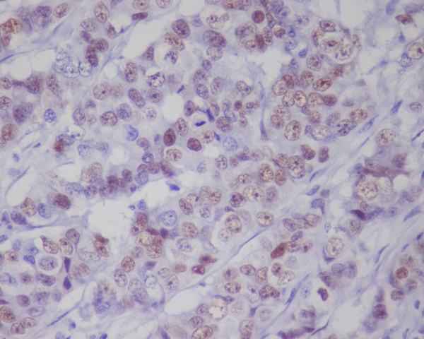
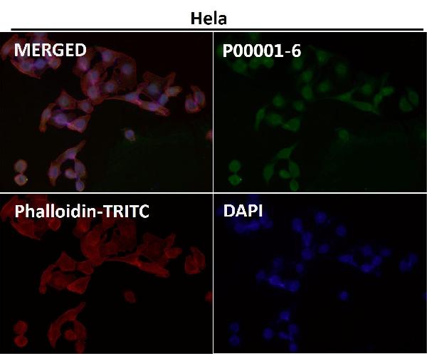
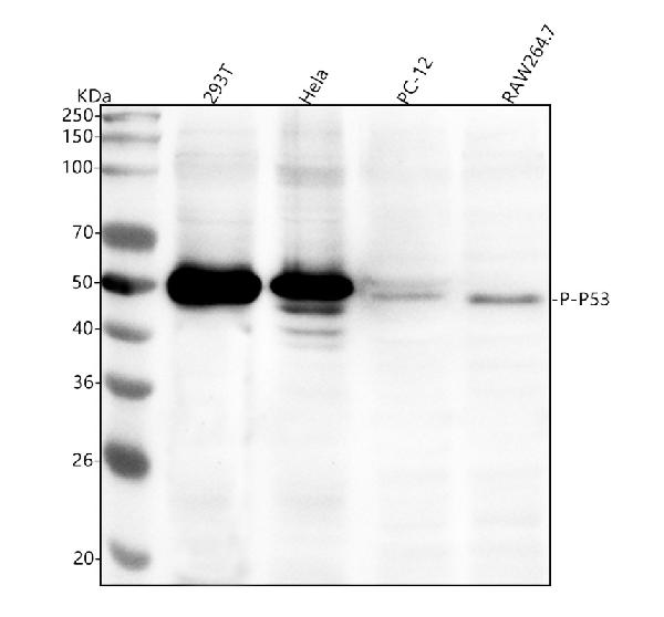
| WB, IHC, IF, ICC, IP |
|---|---|
| Primary Accession | P04637 |
| Host | Rabbit |
| Isotype | Rabbit IgG |
| Reactivity | Rat, Human, Mouse |
| Clonality | Monoclonal |
| Format | Liquid |
| Description | Anti-Phospho-p53 (S392) TP53 Rabbit Monoclonal Antibody . Tested in WB, IHC, ICC/IF, IP applications. This antibody reacts with Human, Mouse, Rat. |
| Gene ID | 7157 |
|---|---|
| Other Names | Cellular tumor antigen p53, Antigen NY-CO-13, Phosphoprotein p53, Tumor suppressor p53, TP53, P53 |
| Calculated MW | 43653 MW KDa |
| Application Details | WB 1:500-1:2000 IHC 1:50-1:200 ICC/IF 1:50-1:200 IP 1:50 |
| Subcellular Localization | Cytoplasm. Nucleus. Nucleus, PML body. Endoplasmic reticulum. Mitochondrion matrix. Interaction with BANP promotes nuclear localization. Recruited into PML bodies together with CHEK2. Translocates to mitochondria upon oxidative stress. |
| Tissue Specificity | Ubiquitous. Isoforms are expressed in a wide range of normal tissues but in a tissue-dependent manner. Isoform 2 is expressed in most normal tissues but is not detected in brain, lung, prostate, muscle, fetal brain, spinal cord and fetal liver. Isoform 3 is expressed in most normal tissues but is not detected in lung, spleen, testis, fetal brain, spinal cord and fetal liver. Isoform 7 is expressed in most normal tissues but is not detected in prostate, uterus, skeletal muscle and breast. Isoform 8 is detected only in colon, bone marrow, testis, fetal brain and intestine. Isoform 9 is expressed in most normal tissues but is not detected in brain, heart, lung, fetal liver, salivary gland, breast or intestine.. |
| Contents | Rabbit IgG in phosphate buffered saline, pH 7.4, 150mM NaCl, 0.02% sodium azide and 50% glycerol, 0.4-0.5mg/ml BSA. |
| Clone Names | Clone: BDO-20 |
| Immunogen | A synthesized peptide derived from human Phospho-p53 (S392) |
| Purification | Affinity-chromatography |
| Storage | Store at -20°C for one year. For short term storage and frequent use, store at 4°C for up to one month. Avoid repeated freeze-thaw cycles. |
| Name | TP53 |
|---|---|
| Synonyms | P53 |
| Function | Multifunctional transcription factor that induces cell cycle arrest, DNA repair or apoptosis upon binding to its target DNA sequence (PubMed:11025664, PubMed:12524540, PubMed:12810724, PubMed:15186775, PubMed:15340061, PubMed:17317671, PubMed:17349958, PubMed:19556538, PubMed:20673990, PubMed:20959462, PubMed:22726440, PubMed:24051492, PubMed:24652652, PubMed:35618207, PubMed:36634798, PubMed:38653238, PubMed:9840937). Acts as a tumor suppressor in many tumor types; induces growth arrest or apoptosis depending on the physiological circumstances and cell type (PubMed:11025664, PubMed:12524540, PubMed:12810724, PubMed:15186775, PubMed:15340061, PubMed:17189187, PubMed:17317671, PubMed:17349958, PubMed:19556538, PubMed:20673990, PubMed:20959462, PubMed:22726440, PubMed:24051492, PubMed:24652652, PubMed:38653238, PubMed:9840937). Negatively regulates cell division by controlling expression of a set of genes required for this process (PubMed:11025664, PubMed:12524540, PubMed:12810724, PubMed:15186775, PubMed:15340061, PubMed:17317671, PubMed:17349958, PubMed:19556538, PubMed:20673990, PubMed:20959462, PubMed:22726440, PubMed:24051492, PubMed:24652652, PubMed:9840937). One of the activated genes is an inhibitor of cyclin-dependent kinases. Apoptosis induction seems to be mediated either by stimulation of BAX and FAS antigen expression, or by repression of Bcl-2 expression (PubMed:12524540, PubMed:17189187). Its pro-apoptotic activity is activated via its interaction with PPP1R13B/ASPP1 or TP53BP2/ASPP2 (PubMed:12524540). However, this activity is inhibited when the interaction with PPP1R13B/ASPP1 or TP53BP2/ASPP2 is displaced by PPP1R13L/iASPP (PubMed:12524540). In cooperation with mitochondrial PPIF is involved in activating oxidative stress-induced necrosis; the function is largely independent of transcription. Induces the transcription of long intergenic non-coding RNA p21 (lincRNA-p21) and lincRNA-Mkln1. LincRNA-p21 participates in TP53-dependent transcriptional repression leading to apoptosis and seems to have an effect on cell-cycle regulation. Implicated in Notch signaling cross-over. Prevents CDK7 kinase activity when associated to CAK complex in response to DNA damage, thus stopping cell cycle progression. Isoform 2 enhances the transactivation activity of isoform 1 from some but not all TP53-inducible promoters. Isoform 4 suppresses transactivation activity and impairs growth suppression mediated by isoform 1. Isoform 7 inhibits isoform 1-mediated apoptosis. Regulates the circadian clock by repressing CLOCK-BMAL1-mediated transcriptional activation of PER2 (PubMed:24051492). |
| Cellular Location | Cytoplasm. Nucleus. Nucleus, PML body. Endoplasmic reticulum. Mitochondrion matrix. Cytoplasm, cytoskeleton, microtubule organizing center, centrosome Note=Recruited into PML bodies together with CHEK2 (PubMed:12810724) Translocates to mitochondria upon oxidative stress (PubMed:22726440) Translocates to mitochondria in response to mitomycin C treatment (PubMed:27323408). Competitive inhibition of TP53 interaction with HSPA9/MOT-2 by UBXN2A results in increased protein abundance and subsequent translocation of TP53 to the nucleus (PubMed:24625977) [Isoform 2]: Nucleus. Cytoplasm. Note=Localized mainly in the nucleus with minor staining in the cytoplasm [Isoform 4]: Nucleus. Cytoplasm. Note=Predominantly nuclear but translocates to the cytoplasm following cell stress [Isoform 8]: Nucleus. Cytoplasm. Note=Localized in both nucleus and cytoplasm in most cells. In some cells, forms foci in the nucleus that are different from nucleoli |
| Tissue Location | Ubiquitous. Isoforms are expressed in a wide range of normal tissues but in a tissue-dependent manner. Isoform 2 is expressed in most normal tissues but is not detected in brain, lung, prostate, muscle, fetal brain, spinal cord and fetal liver. Isoform 3 is expressed in most normal tissues but is not detected in lung, spleen, testis, fetal brain, spinal cord and fetal liver. Isoform 7 is expressed in most normal tissues but is not detected in prostate, uterus, skeletal muscle and breast. Isoform 8 is detected only in colon, bone marrow, testis, fetal brain and intestine. Isoform 9 is expressed in most normal tissues but is not detected in brain, heart, lung, fetal liver, salivary gland, breast or intestine |
Research Areas
Citations (0)

Thousands of laboratories across the world have published research that depended on the performance of antibodies from Abcepta to advance their research. Check out links to articles that cite our products in major peer-reviewed journals, organized by research category.
Submit your citation using an Abcepta antibody to
info@abcepta.com, and receive a free "I Love Antibodies" mug.
info@abcepta.com, and receive a free "I Love Antibodies" mug.

Application Protocols
Provided below are standard protocols that you may find useful for product applications.
Abcepta welcomes feedback from its customers.
If you have used an Abcepta product and would like to share how it has performed, please click on the "Submit Review" button and provide the requested information. Our staff will examine and post your review and contact you if needed.
If you have any additional inquiries please email technical services at tech@abcepta.com.
$ 370.00
Cat# ABO13108
Ordering Information
United States
AlbaniaAustraliaAustriaBelgiumBosnia & HerzegovinaBrazilBulgariaCanadaCentral AmericaChinaCroatiaCyprusCzech RepublicDenmarkEstoniaFinlandFranceGermanyGreeceHong KongHungaryIcelandIndiaIndonesiaIrelandIsraelItalyJapanLatviaLithuaniaLuxembourgMacedoniaMalaysiaMaltaNetherlandsNew ZealandNorwayPakistanPolandPortugalRomaniaSerbiaSingaporeSlovakiaSloveniaSouth AfricaSouth KoreaSpainSwedenSwitzerlandTaiwanTurkeyUnited KingdomUnited StatesVietnamWorldwideOthersMexico
USA Headquarters
(888) 735-7227 / (858) 622-0099 or (858) 875-1900
Other Products
Shipping Information
Domestic orders (in stock items)
Shipped out the same day. Orders placed after 1 PM (PST) will ship out the next business day.
International orders
Contact your local distributors
