


 Foundational characteristics of cancer include proliferation, angiogenesis, migration, evasion of apoptosis, and cellular immortality. Find key markers for these cellular processes and antibodies to detect them.
 The SUMOplot™ Analysis Program predicts and scores sumoylation sites in your protein. SUMOylation is a post-translational modification involved in various cellular processes, such as nuclear-cytosolic transport, transcriptional regulation, apoptosis, protein stability, response to stress, and progression through the cell cycle.
 The Autophagy Receptor Motif Plotter predicts and scores autophagy receptor binding sites in your protein. Identifying proteins connected to this pathway is critical to understanding the role of autophagy in physiological as well as pathological processes such as development, differentiation, neurodegenerative diseases, stress, infection, and cancer.
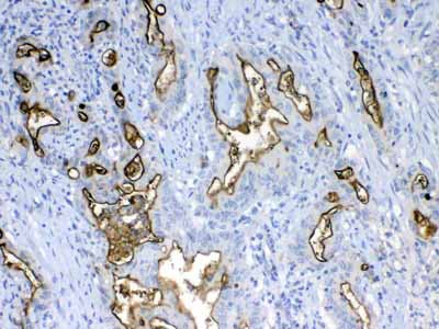
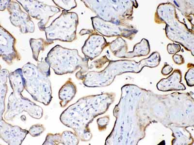
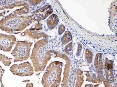
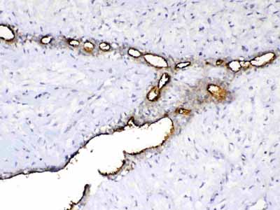
Anti-MUC1 Picoband Antibody
- SPECIFICATION
- CITATIONS
- PROTOCOLS
- BACKGROUND

Application
| WB, IHC-P, E |
|---|---|
| Primary Accession | P15941 |
| Host | Rabbit |
| Reactivity | Human, Mouse, Rat |
| Clonality | Polyclonal |
| Format | Lyophilized |
| Description | Rabbit IgG polyclonal antibody for MUC1 detection. Tested with WB, IHC-P, Direct ELISA in Human;Mouse;Rat. |
| Reconstitution | Add 0.2ml of distilled water will yield a concentration of 500ug/ml. |
| Gene ID | 4582 |
|---|---|
| Other Names | Mucin-1, MUC-1, Breast carcinoma-associated antigen DF3, Cancer antigen 15-3, CA 15-3, Carcinoma-associated mucin, Episialin, H23AG, Krebs von den Lungen-6, KL-6, PEMT, Peanut-reactive urinary mucin, PUM, Polymorphic epithelial mucin, PEM, Tumor-associated epithelial membrane antigen, EMA, Tumor-associated mucin, CD227, Mucin-1 subunit alpha, MUC1-NT, MUC1-alpha, Mucin-1 subunit beta, MUC1-beta, MUC1-CT, MUC1, PUM |
| Calculated MW | 122102 Da |
| Application Details | Western blot, 0.1-0.5 µg/ml Immunohistochemistry(Paraffin-embedded Section), 0.5-1 µg/ml Direct ELISA, 0.1-0.5 µg/ml |
| Subcellular Localization | Apical cell membrane. |
| Tissue Specificity | Expressed on the apical surface of epithelial cells, especially of airway passages, breast and uterus. Also expressed in activated and unactivated T-cells. Overexpressed in epithelial tumors, such as breast or ovarian cancer and also in non-epithelial tumor cells. Isoform Y is expressed in tumor cells only. |
| Contents | Each vial contains 4mg Trehalose, 0.9mg NaCl, 0.2mg Na2HPO4, 0.05mg NaN3. |
| Immunogen | E. coli-derived human MUC1 recombinant protein (Position: S1062-V1139). |
| Cross Reactivity | No cross reactivity with other proteins. |
| Storage | At -20˚C; for one year. After r˚Constitution, at 4˚C; for one month. It˚Can also be aliquotted and stored frozen at -20˚C; for a longer time. Avoid repeated freezing and thawing. |
| Name | MUC1 |
|---|---|
| Synonyms | PUM |
| Function | The alpha subunit has cell adhesive properties. Can act both as an adhesion and an anti-adhesion protein. May provide a protective layer on epithelial cells against bacterial and enzyme attack. |
| Cellular Location | Apical cell membrane; Single-pass type I membrane protein. Note=Exclusively located in the apical domain of the plasma membrane of highly polarized epithelial cells After endocytosis, internalized and recycled to the cell membrane Located to microvilli and to the tips of long filopodial protusions [Isoform Y]: Secreted. [Mucin-1 subunit beta]: Cell membrane. Cytoplasm. Nucleus. Note=On EGF and PDGFRB stimulation, transported to the nucleus through interaction with CTNNB1, a process which is stimulated by phosphorylation. On HRG stimulation, colocalizes with JUP/gamma-catenin at the nucleus |
| Tissue Location | Expressed on the apical surface of epithelial cells, especially of airway passages, breast and uterus. Also expressed in activated and unactivated T-cells. Overexpressed in epithelial tumors, such as breast or ovarian cancer and also in non-epithelial tumor cells. Isoform Y is expressed in tumor cells only |

Thousands of laboratories across the world have published research that depended on the performance of antibodies from Abcepta to advance their research. Check out links to articles that cite our products in major peer-reviewed journals, organized by research category.
info@abcepta.com, and receive a free "I Love Antibodies" mug.

Provided below are standard protocols that you may find useful for product applications.
Background
Mucin 1, cell surface associated (MUC1) or polymorphic epithelial mucin (PEM) is a mucin encoded by the MUC1 gene in humans. This gene encodes a membrane-bound protein that is a member of the mucin family. Mucins are O-glycosylated proteins that play an essential role in forming protective mucous barriers on epithelial surfaces. It is mapped to 1q22. Mucin 1 is a transmembrane mucin normally expressed on the apical borders of secretory epithelial cells. Overexpression of Mucin 1 is often associated with colon, breast, ovarian, lung and pancreatic cancers. The protein serves a protective function by binding to pathogens and also functions in a cell signaling capacity. Mucin 1 stimulated ESR1-mediated transcription and contributed to estradiol-mediated growth and survival of breast cancer cells. This gene also can suppress pulmonary innate immunity, and its antiinflammatory activity may play an important modulatory role during microbial infection.
If you have used an Abcepta product and would like to share how it has performed, please click on the "Submit Review" button and provide the requested information. Our staff will examine and post your review and contact you if needed.
If you have any additional inquiries please email technical services at tech@abcepta.com.
Ordering Information
Other Products
Shipping Information
