


 Foundational characteristics of cancer include proliferation, angiogenesis, migration, evasion of apoptosis, and cellular immortality. Find key markers for these cellular processes and antibodies to detect them.
 The SUMOplot™ Analysis Program predicts and scores sumoylation sites in your protein. SUMOylation is a post-translational modification involved in various cellular processes, such as nuclear-cytosolic transport, transcriptional regulation, apoptosis, protein stability, response to stress, and progression through the cell cycle.
 The Autophagy Receptor Motif Plotter predicts and scores autophagy receptor binding sites in your protein. Identifying proteins connected to this pathway is critical to understanding the role of autophagy in physiological as well as pathological processes such as development, differentiation, neurodegenerative diseases, stress, infection, and cancer.
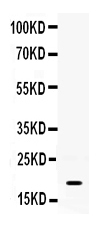
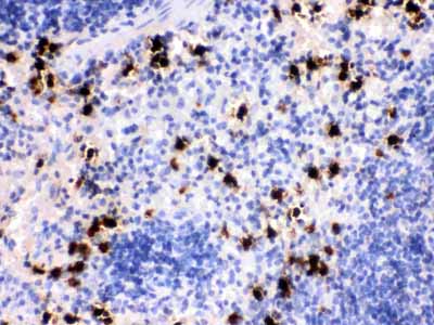
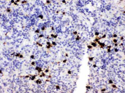
Anti-S100A8 Antibody
- SPECIFICATION
- CITATIONS
- PROTOCOLS
- BACKGROUND

Application
| WB, IHC-P |
|---|---|
| Primary Accession | P27005 |
| Host | Rabbit |
| Reactivity | Mouse, Rat |
| Clonality | Polyclonal |
| Format | Lyophilized |
| Description | Rabbit IgG polyclonal antibody for Protein S100-A8(S100A8) detection. Tested with WB, IHC-P in Mouse;Rat. |
| Reconstitution | Add 0.2ml of distilled water will yield a concentration of 500ug/ml. |
| Gene ID | 20201 |
|---|---|
| Other Names | Protein S100-A8, Calgranulin-A, Chemotactic cytokine CP-10, Leukocyte L1 complex light chain, Migration inhibitory factor-related protein 8, MRP-8, p8, Pro-inflammatory S100 cytokine, S100 calcium-binding protein A8, S100a8, Caga, Mrp8 |
| Calculated MW | 10295 MW KDa |
| Application Details | Immunohistochemistry(Paraffin-embedded Section), 0.5-1 µg/ml, Mouse, Rat, By Heat Western blot, 0.1-0.5 µg/ml, Mouse |
| Subcellular Localization | Secreted . Cytoplasm . Cytoplasm, cytoskeleton . Cell membrane ; Peripheral membrane protein . Predominantly localized in the cytoplasm. Upon elevation of the intracellular calcium level, translocated from the cytoplasm to the cytoskeleton and the cell membrane. Upon neutrophil activation or endothelial adhesion of monocytes, is secreted via a microtubule-mediated, alternative pathway. |
| Protein Name | Protein S100-A8 |
| Contents | Each vial contains 5mg BSA, 0.9mg NaCl, 0.2mg Na2HPO4, 0.05mg NaN3. |
| Immunogen | E.coli-derived mouse S100A8 recombinant protein (Position: P2-E89). Mouse S100A8 shares 58.6% and 80.5% amino acid (aa) sequence identity with human and rat S100A8, respectively. |
| Purification | Immunogen affinity purified. |
| Cross Reactivity | No cross reactivity with other proteins. |
| Storage | At -20˚C for one year. After r˚Constitution, at 4˚C for one month. It˚Can also be aliquotted and stored frozen at -20˚C for a longer time.Avoid repeated freezing and thawing. |
| Name | S100a8 {ECO:0000312|MGI:MGI:88244} |
|---|---|
| Synonyms | Caga, Mrp8 |
| Function | S100A8 is a calcium- and zinc-binding protein which plays a prominent role in the regulation of inflammatory processes and immune response. It can induce neutrophil chemotaxis and adhesion. Predominantly found as calprotectin (S100A8/A9) which has a wide plethora of intra- and extracellular functions. The intracellular functions include: facilitating leukocyte arachidonic acid trafficking and metabolism, modulation of the tubulin-dependent cytoskeleton during migration of phagocytes and activation of the neutrophilic NADPH- oxidase. Participates also in regulatory T-cell differentiation together with CD69. Activates NADPH-oxidase by facilitating the enzyme complex assembly at the cell membrane, transferring arachidonic acid, an essential cofactor, to the enzyme complex and S100A8 contributes to the enzyme assembly by directly binding to NCF2/P67PHOX. The extracellular functions involve pro-inflammatory, antimicrobial, oxidant-scavenging and apoptosis-inducing activities. Its pro- inflammatory activity includes recruitment of leukocytes, promotion of cytokine and chemokine production, and regulation of leukocyte adhesion and migration. Acts as an alarmin or a danger associated molecular pattern (DAMP) molecule and stimulates innate immune cells via binding to pattern recognition receptors such as Toll-like receptor 4 (TLR4) and receptor for advanced glycation endproducts (AGER). Binding to TLR4 and AGER activates the MAP-kinase and NF-kappa-B signaling pathways resulting in the amplification of the pro-inflammatory cascade. Has antimicrobial activity towards bacteria and fungi and exerts its antimicrobial activity probably via chelation of Zn(2+) which is essential for microbial growth. Can induce cell death via autophagy and apoptosis and this occurs through the cross-talk of mitochondria and lysosomes via reactive oxygen species (ROS) and the process involves BNIP3. Can regulate neutrophil number and apoptosis by an anti- apoptotic effect; regulates cell survival via ITGAM/ITGB and TLR4 and a signaling mechanism involving MEK-ERK. Its role as an oxidant scavenger has a protective role in preventing exaggerated tissue damage by scavenging oxidants. The iNOS-S100A8/A9 transnitrosylase complex is proposed to direct selective inflammatory stimulus-dependent S- nitrosylation of multiple targets such as GAPDH, ANXA5, EZR, MSN and VIM by recognizing a [IL]-x-C-x-x-[DE] motif; S100A8 seems to contribute to S-nitrosylation site selectivity (By similarity). |
| Cellular Location | Secreted. Cytoplasm. Cytoplasm, cytoskeleton. Cell membrane; Peripheral membrane protein Note=Predominantly localized in the cytoplasm. Upon elevation of the intracellular calcium level, translocated from the cytoplasm to the cytoskeleton and the cell membrane. Upon neutrophil activation or endothelial adhesion of monocytes, is secreted via a microtubule- mediated, alternative pathway |

Thousands of laboratories across the world have published research that depended on the performance of antibodies from Abcepta to advance their research. Check out links to articles that cite our products in major peer-reviewed journals, organized by research category.
info@abcepta.com, and receive a free "I Love Antibodies" mug.

Provided below are standard protocols that you may find useful for product applications.
Background
S100 calcium-binding protein A8 (S100A8) is a protein that in humans is encoded by the S100A8 gene. It is also known as calgranulin A. The protein encoded by this gene is a member of the S100 family of proteins containing 2 EF-hand calcium-binding motifs. S100 proteins are localized in the cytoplasm and/or nucleus of a wide range of cells, and involved in the regulation of a number of cellular processes such as cell cycle progression and differentiation. S100 genes include at least 13 members which are located as a cluster on chromosome 1q21. This protein may function in the inhibition of casein kinase and as a cytokine. Altered expression of this protein is associated with the disease cystic fibrosis.
If you have used an Abcepta product and would like to share how it has performed, please click on the "Submit Review" button and provide the requested information. Our staff will examine and post your review and contact you if needed.
If you have any additional inquiries please email technical services at tech@abcepta.com.
Ordering Information
Shipping Information
