


 Foundational characteristics of cancer include proliferation, angiogenesis, migration, evasion of apoptosis, and cellular immortality. Find key markers for these cellular processes and antibodies to detect them.
 The SUMOplot™ Analysis Program predicts and scores sumoylation sites in your protein. SUMOylation is a post-translational modification involved in various cellular processes, such as nuclear-cytosolic transport, transcriptional regulation, apoptosis, protein stability, response to stress, and progression through the cell cycle.
 The Autophagy Receptor Motif Plotter predicts and scores autophagy receptor binding sites in your protein. Identifying proteins connected to this pathway is critical to understanding the role of autophagy in physiological as well as pathological processes such as development, differentiation, neurodegenerative diseases, stress, infection, and cancer.
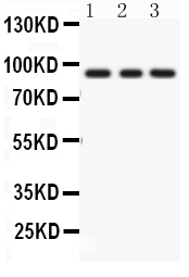
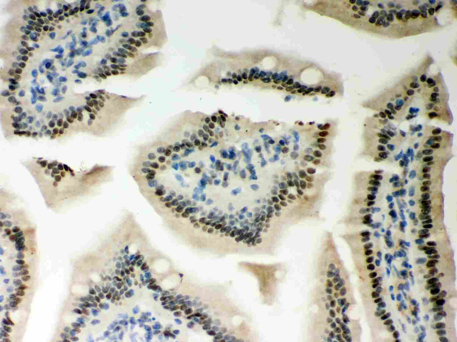
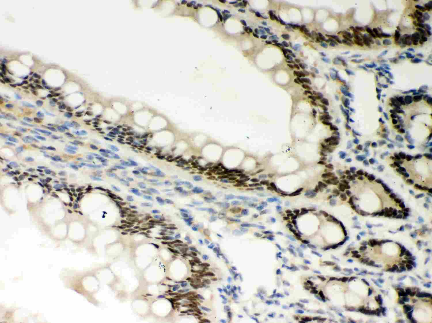
Anti-PKC Epsilon Picoband Antibody
- SPECIFICATION
- CITATIONS
- PROTOCOLS
- BACKGROUND

Application
| WB, IHC-P |
|---|---|
| Primary Accession | Q02156 |
| Host | Rabbit |
| Reactivity | Human, Mouse, Rat |
| Clonality | Polyclonal |
| Format | Lyophilized |
| Description | Rabbit IgG polyclonal antibody for Protein kinase C epsilon type(PRKCE) detection. Tested with WB, IHC-P in Human;Mouse;Rat. |
| Reconstitution | Add 0.2ml of distilled water will yield a concentration of 500ug/ml. |
| Gene ID | 5581 |
|---|---|
| Other Names | Protein kinase C epsilon type, 2.7.11.13, nPKC-epsilon, PRKCE, PKCE |
| Calculated MW | 83674 MW KDa |
| Application Details | Immunohistochemistry(Paraffin-embedded Section), 0.5-1 µg/ml, Mouse, Rat, Human, By Heat Western blot, 0.1-0.5 µg/ml, Human, Mouse, Rat |
| Subcellular Localization | Cytoplasm . Cytoplasm, cytoskeleton . Cell membrane . Cytoplasm, perinuclear region . Nucleus . Translocated to plasma membrane in epithelial cells stimulated by HGF. Associated with the Golgi at the perinuclear site in pre-passage fibroblasts (By similarity). In passaging cells, translocated to the cell periphery (By similarity). Translocated to the nucleus in PMA- treated cells (By similarity). . |
| Protein Name | Protein kinase C epsilon type |
| Contents | Each vial contains 5mg BSA, 0.9mg NaCl, 0.2mg Na2HPO4, 0.05mg NaN3. |
| Immunogen | E.coli-derived human PKC epsilon recombinant protein (Position: Q53-R236). Human PKC epsilon shares 99% amino acid (aa) sequence identity with both mouse and rat PKC epsilon. |
| Purification | Immunogen affinity purified. |
| Cross Reactivity | No cross reactivity with other proteins |
| Storage | At -20˚C for one year. After r˚Constitution, at 4˚C for one month. It˚Can also be aliquotted and stored frozen at -20˚C for a longer time.Avoid repeated freezing and thawing. |
| Sequence Similarities | Belongs to the protein kinase superfamily. AGC Ser/Thr protein kinase family. PKC subfamily. |
| Name | PRKCE |
|---|---|
| Synonyms | PKCE |
| Function | Calcium-independent, phospholipid- and diacylglycerol (DAG)- dependent serine/threonine-protein kinase that plays essential roles in the regulation of multiple cellular processes linked to cytoskeletal proteins, such as cell adhesion, motility, migration and cell cycle, functions in neuron growth and ion channel regulation, and is involved in immune response, cancer cell invasion and regulation of apoptosis. Mediates cell adhesion to the extracellular matrix via integrin- dependent signaling, by mediating angiotensin-2-induced activation of integrin beta-1 (ITGB1) in cardiac fibroblasts. Phosphorylates MARCKS, which phosphorylates and activates PTK2/FAK, leading to the spread of cardiomyocytes. Involved in the control of the directional transport of ITGB1 in mesenchymal cells by phosphorylating vimentin (VIM), an intermediate filament (IF) protein. In epithelial cells, associates with and phosphorylates keratin-8 (KRT8), which induces targeting of desmoplakin at desmosomes and regulates cell-cell contact. Phosphorylates IQGAP1, which binds to CDC42, mediating epithelial cell- cell detachment prior to migration. In HeLa cells, contributes to hepatocyte growth factor (HGF)-induced cell migration, and in human corneal epithelial cells, plays a critical role in wound healing after activation by HGF. During cytokinesis, forms a complex with YWHAB, which is crucial for daughter cell separation, and facilitates abscission by a mechanism which may implicate the regulation of RHOA. In cardiac myocytes, regulates myofilament function and excitation coupling at the Z-lines, where it is indirectly associated with F-actin via interaction with COPB1. During endothelin-induced cardiomyocyte hypertrophy, mediates activation of PTK2/FAK, which is critical for cardiomyocyte survival and regulation of sarcomere length. Plays a role in the pathogenesis of dilated cardiomyopathy via persistent phosphorylation of troponin I (TNNI3). Involved in nerve growth factor (NFG)-induced neurite outgrowth and neuron morphological change independently of its kinase activity, by inhibition of RHOA pathway, activation of CDC42 and cytoskeletal rearrangement. May be involved in presynaptic facilitation by mediating phorbol ester-induced synaptic potentiation. Phosphorylates gamma-aminobutyric acid receptor subunit gamma-2 (GABRG2), which reduces the response of GABA receptors to ethanol and benzodiazepines and may mediate acute tolerance to the intoxicating effects of ethanol. Upon PMA treatment, phosphorylates the capsaicin- and heat-activated cation channel TRPV1, which is required for bradykinin-induced sensitization of the heat response in nociceptive neurons. Is able to form a complex with PDLIM5 and N-type calcium channel, and may enhance channel activities and potentiates fast synaptic transmission by phosphorylating the pore-forming alpha subunit CACNA1B (CaV2.2). In prostate cancer cells, interacts with and phosphorylates STAT3, which increases DNA-binding and transcriptional activity of STAT3 and seems to be essential for prostate cancer cell invasion. Downstream of TLR4, plays an important role in the lipopolysaccharide (LPS)-induced immune response by phosphorylating and activating TICAM2/TRAM, which in turn activates the transcription factor IRF3 and subsequent cytokines production. In differentiating erythroid progenitors, is regulated by EPO and controls the protection against the TNFSF10/TRAIL-mediated apoptosis, via BCL2. May be involved in the regulation of the insulin-induced phosphorylation and activation of AKT1. Phosphorylates NLRP5/MATER and may thereby modulate AKT pathway activation in cumulus cells (PubMed:19542546). Phosphorylates and activates LRRK1, which phosphorylates RAB proteins involved in intracellular trafficking (PubMed:36040231). |
| Cellular Location | Cytoplasm. Cytoplasm, cytoskeleton. Cell membrane. Cytoplasm, perinuclear region {ECO:0000250|UniProtKB:P16054}. Nucleus {ECO:0000250|UniProtKB:P16054} Note=Translocated to plasma membrane in epithelial cells stimulated by HGF (PubMed:17603037). Associated with the Golgi at the perinuclear site in pre-passage fibroblasts (By similarity). In passaging cells, translocated to the cell periphery (By similarity). Translocated to the nucleus in PMA-treated cells (By similarity) {ECO:0000250|UniProtKB:P16054, ECO:0000269|PubMed:17603037} |
| Tissue Location | Expressed in cumulus cells (at protein level). |

Thousands of laboratories across the world have published research that depended on the performance of antibodies from Abcepta to advance their research. Check out links to articles that cite our products in major peer-reviewed journals, organized by research category.
info@abcepta.com, and receive a free "I Love Antibodies" mug.

Provided below are standard protocols that you may find useful for product applications.
Background
Protein kinase C epsilon type, also known as PKCE, is an enzyme that in humans is encoded by the PRKCE gene. The protein encoded by this gene is one of the PKC family members. PRKCE is mapped to 2p21. This kinase has been shown to be involved in many different cellular functions, such as apoptosis, cardioprotection from ischemia, heat shock response, as well as insulin exocytosis. It has been found that activation of PRKCE can induce VR1 channel activity at room temperature in the absence of any other agonist. PRKCE gene plays a role in apoptosis signaling pathways in thyroid cells and it has been indicated that a naturally occurring PRKCE mutant that functions as a dominant negative can block cell death triggered by a variety of stimuli. Expression of PRKCE inhibits chemotherapy-induced caspase-3 activation and apoptosis, thereby leading to cell survival.
If you have used an Abcepta product and would like to share how it has performed, please click on the "Submit Review" button and provide the requested information. Our staff will examine and post your review and contact you if needed.
If you have any additional inquiries please email technical services at tech@abcepta.com.
Ordering Information
Other Products
Shipping Information
