Anti-ALK Antibody
- SPECIFICATION
- CITATIONS
- PROTOCOLS
- BACKGROUND

Application
| WB, IHC-P |
|---|---|
| Primary Accession | Q9UM73 |
| Host | Rabbit |
| Reactivity | Human, Mouse, Rat |
| Clonality | Polyclonal |
| Format | Lyophilized |
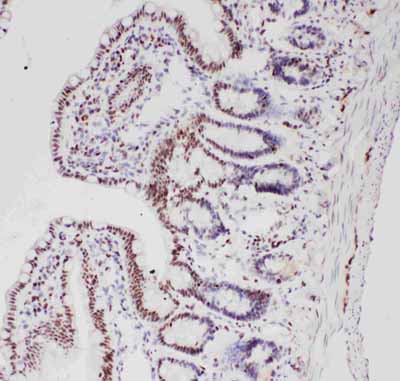
| Description | Rabbit IgG polyclonal antibody for ALK tyrosine kinase receptor(ALK) detection. Tested with WB, IHC-P in Human;Mouse;Rat. |
| Reconstitution | Add 0.2ml of distilled water will yield a concentration of 500ug/ml. |
| Gene ID | 238 |
|---|---|
| Other Names | ALK tyrosine kinase receptor, 2.7.10.1, Anaplastic lymphoma kinase, CD246, ALK |
| Calculated MW | 176442 MW KDa |
| Application Details | Immunohistochemistry(Paraffin-embedded Section), 0.5-1 µg/ml, Human, Rat, Mouse, By Heat Western blot, 0.1-0.5 µg/ml, Human, Rat, Mouse |
| Subcellular Localization | Cell membrane ; Single-pass type I membrane protein . Membrane attachment was crucial for promotion of neuron-like differentiation and cell proliferation arrest through specific activation of the MAP kinase pathway. |
| Tissue Specificity | Expressed in brain and CNS. Also expressed in the small intestine and testis, but not in normal lymphoid cells. . |
| Protein Name | ALK tyrosine kinase receptor |
| Contents | Each vial contains 5mg BSA, 0.9mg NaCl, 0.2mg Na2HPO4, 0.05mg Thimerosal, 0.05mg NaN3. |
| Immunogen | A synthetic peptide corresponding to a sequence at the C-terminus of human ALK(1571-1589aa LFRLRHFPCGNVNYGYQQQ), identical to the related rat and mouse sequences. |
| Purification | Immunogen affinity purified. |
| Cross Reactivity | No cross reactivity with other proteins |
| Storage | At -20˚C for one year. After r˚Constitution, at 4˚C for one month. It˚Can also be aliquotted and stored frozen at -20˚C for a longer time.Avoid repeated freezing and thawing. |
| Sequence Similarities | Belongs to the protein kinase superfamily. Tyr protein kinase family. Insulin receptor subfamily. |
| Name | ALK {ECO:0000303|PubMed:9174053, ECO:0000312|HGNC:HGNC:427} |
|---|---|
| Function | Neuronal receptor tyrosine kinase that is essentially and transiently expressed in specific regions of the central and peripheral nervous systems and plays an important role in the genesis and differentiation of the nervous system (PubMed:11121404, PubMed:11387242, PubMed:16317043, PubMed:17274988, PubMed:30061385, PubMed:34646012, PubMed:34819673). Also acts as a key thinness protein involved in the resistance to weight gain: in hypothalamic neurons, controls energy expenditure acting as a negative regulator of white adipose tissue lipolysis and sympathetic tone to fine-tune energy homeostasis (By similarity). Following activation by ALKAL2 ligand at the cell surface, transduces an extracellular signal into an intracellular response (PubMed:30061385, PubMed:33411331, PubMed:34646012, PubMed:34819673). In contrast, ALKAL1 is not a potent physiological ligand for ALK (PubMed:34646012). Ligand-binding to the extracellular domain induces tyrosine kinase activation, leading to activation of the mitogen-activated protein kinase (MAPK) pathway (PubMed:34819673). Phosphorylates almost exclusively at the first tyrosine of the Y-x-x-x-Y-Y motif (PubMed:15226403, PubMed:16878150). Induces tyrosine phosphorylation of CBL, FRS2, IRS1 and SHC1, as well as of the MAP kinases MAPK1/ERK2 and MAPK3/ERK1 (PubMed:15226403, PubMed:16878150). ALK activation may also be regulated by pleiotrophin (PTN) and midkine (MDK) (PubMed:11278720, PubMed:11809760, PubMed:12107166, PubMed:12122009). PTN-binding induces MAPK pathway activation, which is important for the anti-apoptotic signaling of PTN and regulation of cell proliferation (PubMed:11278720, PubMed:11809760, PubMed:12107166). MDK-binding induces phosphorylation of the ALK target insulin receptor substrate (IRS1), activates mitogen-activated protein kinases (MAPKs) and PI3-kinase, resulting also in cell proliferation induction (PubMed:12122009). Drives NF-kappa-B activation, probably through IRS1 and the activation of the AKT serine/threonine kinase (PubMed:15226403, PubMed:16878150). Recruitment of IRS1 to activated ALK and the activation of NF-kappa-B are essential for the autocrine growth and survival signaling of MDK (PubMed:15226403, PubMed:16878150). |
| Cellular Location | Cell membrane; Single-pass type I membrane protein Note=Membrane attachment is essential for promotion of neuron-like differentiation and cell proliferation arrest through specific activation of the MAP kinase pathway. |
| Tissue Location | Expressed in brain and CNS. Also expressed in the small intestine and testis, but not in normal lymphoid cells |

Thousands of laboratories across the world have published research that depended on the performance of antibodies from Abcepta to advance their research. Check out links to articles that cite our products in major peer-reviewed journals, organized by research category.
info@abcepta.com, and receive a free "I Love Antibodies" mug.
Provided below are standard protocols that you may find useful for product applications.
Background
ALK(Anaplastic lymphoma kinase) also known as ALK tyrosine kinase receptor or CD246(cluster of differentiation 246) is an enzyme that in humans is encoded by the ALK gene. The ALK gene is mapped on 2p23.2-p23.1. Expressed in the small intestine, testis, and brain but not in normal lymphoid cells, ALK shows greatest sequence similarity to the insulin receptor subfamily of kinases. ALK plays an important role in the development of the brain and exerts its effects on specific neurons in the nervous system. The deduced amino acid sequences reveal that ALK is a novel receptor tyrosine kinase having a putative transmembrane domain and an extracellular domain. In Drosophila, localized Jeb activates Alk and the downstream Ras/mitogen-activated protein kinase cascade to specify a select group of visceral muscle precursors as muscle-patterning pioneers. Functional RNA interference screening on a set of these transcriptional targets revealed that CEBPB and BCL2A1 were absolutely necessary to induce cell transformation and/or to sustain growth and survival of ALK-positive ALCL cells. One particularly informative case presented a high-level gene amplification that was strictly limited to ALK, indicating that this gene may contribute on its own to neuroblastoma development. Mutated ALK proteins were overexpressed, hyperphosphorylated, and showed constitutive kinase activity. The knockdown of ALK expression in ALK-mutated cells, but also in cell lines overexpressing a wildtype ALK, led to a marked decrease of cell proliferation.
If you have used an Abcepta product and would like to share how it has performed, please click on the "Submit Review" button and provide the requested information. Our staff will examine and post your review and contact you if needed.
If you have any additional inquiries please email technical services at tech@abcepta.com.


 Foundational characteristics of cancer include proliferation, angiogenesis, migration, evasion of apoptosis, and cellular immortality. Find key markers for these cellular processes and antibodies to detect them.
 The SUMOplot™ Analysis Program predicts and scores sumoylation sites in your protein. SUMOylation is a post-translational modification involved in various cellular processes, such as nuclear-cytosolic transport, transcriptional regulation, apoptosis, protein stability, response to stress, and progression through the cell cycle.
 The Autophagy Receptor Motif Plotter predicts and scores autophagy receptor binding sites in your protein. Identifying proteins connected to this pathway is critical to understanding the role of autophagy in physiological as well as pathological processes such as development, differentiation, neurodegenerative diseases, stress, infection, and cancer.


