


 Foundational characteristics of cancer include proliferation, angiogenesis, migration, evasion of apoptosis, and cellular immortality. Find key markers for these cellular processes and antibodies to detect them.
 The SUMOplot™ Analysis Program predicts and scores sumoylation sites in your protein. SUMOylation is a post-translational modification involved in various cellular processes, such as nuclear-cytosolic transport, transcriptional regulation, apoptosis, protein stability, response to stress, and progression through the cell cycle.
 The Autophagy Receptor Motif Plotter predicts and scores autophagy receptor binding sites in your protein. Identifying proteins connected to this pathway is critical to understanding the role of autophagy in physiological as well as pathological processes such as development, differentiation, neurodegenerative diseases, stress, infection, and cancer.
Anti-NDRG1 Antibody
- SPECIFICATION
- CITATIONS
- PROTOCOLS
- BACKGROUND

Application
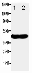
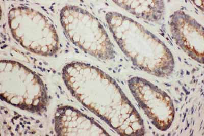
| WB, IHC-P |
|---|---|
| Primary Accession | Q92597 |
| Host | Rabbit |
| Reactivity | Human |
| Clonality | Polyclonal |
| Format | Lyophilized |
| Description | Rabbit IgG polyclonal antibody for Protein NDRG1(NDRG1) detection. Tested with WB, IHC-P in Human. |
| Reconstitution | Add 0.2ml of distilled water will yield a concentration of 500ug/ml. |
| Gene ID | 10397 |
|---|---|
| Other Names | Protein NDRG1, Differentiation-related gene 1 protein, DRG-1, N-myc downstream-regulated gene 1 protein, Nickel-specific induction protein Cap43, Reducing agents and tunicamycin-responsive protein, RTP, Rit42, NDRG1, CAP43, DRG1, RTP |
| Calculated MW | 42835 MW KDa |
| Application Details | Immunohistochemistry(Paraffin-embedded Section), 0.5-1 µg/ml, Human, By Heat Western blot, 0.1-0.5 µg/ml, Human |
| Subcellular Localization | Cytoplasm, cytosol. Cytoplasm, cytoskeleton, microtubule organizing center, centrosome. Nucleus. Cell membrane. Mainly cytoplasmic but differentially localized to other regions. Associates with the plasma membrane in intestinal epithelia and lactating mammary gland. Translocated to the nucleus in a p53/TP53-dependent manner. In prostate epithelium and placental chorion, located in both the cytoplasm and in the nucleus. No nuclear localization in colon epithelium cells. In intestinal mucosa, prostate and renal cortex, located predominantly adjacent to adherens junctions. Cytoplasmic with granular staining in proximal tubular cells of the kidney and salivary gland ducts. Recruits to the membrane of recycling/sorting and late endosomes via binding to phosphatidylinositol 4-phosphate. Associates with microtubules. Colocalizes with TUBG1 in the centrosome. Cytoplasmic location increased with hypoxia. Phosphorylated form found associated with centromeres during S-phase of mitosis and with the plasma membrane. |
| Tissue Specificity | Ubiquitous; expressed most prominently in placental membranes and prostate, kidney, small intestine, and ovary tissues. Also expressed in heart, brain, skeletal muscle, lung, liver and pancreas. Low levels in peripheral blood leukocytes and in tissues of the immune system. Expressed mainly in epithelial cells. Also found in Schwann cells of peripheral neurons. Reduced expression in adenocarcinomas compared to normal tissues. In colon, prostate and placental membranes, the cells that border the lumen show the highest expression. . |
| Protein Name | Protein NDRG1 |
| Contents | Each vial contains 5mg BSA, 0.9mg NaCl, 0.2mg Na2HPO4, 0.05mg Thimerosal, 0.05mg NaN3. |
| Immunogen | A synthetic peptide corresponding to a sequence in the middle region of human NDRG1(194-214aa HLFGKEEMQSNVEVVHTYRQH). |
| Purification | Immunogen affinity purified. |
| Cross Reactivity | No cross reactivity with other proteins |
| Storage | At -20˚C for one year. After r˚Constitution, at 4˚C for one month. It˚Can also be aliquotted and stored frozen at -20˚C for a longer time.Avoid repeated freezing and thawing. |
| Sequence Similarities | Belongs to the NDRG family. |
| Name | NDRG1 |
|---|---|
| Synonyms | CAP43, DRG1, RTP |
| Function | Stress-responsive protein involved in hormone responses, cell growth, and differentiation. Acts as a tumor suppressor in many cell types. Necessary but not sufficient for p53/TP53-mediated caspase activation and apoptosis. Has a role in cell trafficking, notably of the Schwann cell, and is necessary for the maintenance and development of the peripheral nerve myelin sheath. Required for vesicular recycling of CDH1 and TF. May also function in lipid trafficking. Protects cells from spindle disruption damage. Functions in p53/TP53-dependent mitotic spindle checkpoint. Regulates microtubule dynamics and maintains euploidy. |
| Cellular Location | Cytoplasm, cytosol. Cytoplasm, cytoskeleton, microtubule organizing center, centrosome. Nucleus. Cell membrane Note=Mainly cytoplasmic but differentially localized to other regions Associates with the plasma membrane in intestinal epithelia and lactating mammary gland. Translocated to the nucleus in a p53/TP53- dependent manner. In prostate epithelium and placental chorion, located in both the cytoplasm and in the nucleus. No nuclear localization in colon epithelium cells. In intestinal mucosa, prostate and renal cortex, located predominantly adjacent to adherens junctions Cytoplasmic with granular staining in proximal tubular cells of the kidney and salivary gland ducts. Recruits to the membrane of recycling/sorting and late endosomes via binding to phosphatidylinositol 4-phosphate. Associates with microtubules Colocalizes with TUBG1 in the centrosome. Cytoplasmic location increased with hypoxia. Phosphorylated form found associated with centromeres during S-phase of mitosis and with the plasma membrane |
| Tissue Location | Ubiquitous; expressed most prominently in placental membranes and prostate, kidney, small intestine, and ovary tissues Also expressed in heart, brain, skeletal muscle, lung, liver and pancreas. Low levels in peripheral blood leukocytes and in tissues of the immune system. Expressed mainly in epithelial cells. Also found in Schwann cells of peripheral neurons. Reduced expression in adenocarcinomas compared to normal tissues. In colon, prostate and placental membranes, the cells that border the lumen show the highest expression. |

Thousands of laboratories across the world have published research that depended on the performance of antibodies from Abcepta to advance their research. Check out links to articles that cite our products in major peer-reviewed journals, organized by research category.
info@abcepta.com, and receive a free "I Love Antibodies" mug.

Provided below are standard protocols that you may find useful for product applications.
Background
Protein NDRG1 is a protein that in humans is encoded by the NDRG1 gene. It is a member of the N-myc downregulated gene family which belong to the alpha/beta hydrolase superfamily. The NDRG1 gene is mapped to chromosome 8q24. It has got a deduced 394 amino acid protein with a molecular mass of 43kD. NDRG1 is expressed in the cytoplasm and basolateral membranes of gut lumen surface epithelial cells. The protein appears to play a vital role in growth arrest and cell differentiation.
If you have used an Abcepta product and would like to share how it has performed, please click on the "Submit Review" button and provide the requested information. Our staff will examine and post your review and contact you if needed.
If you have any additional inquiries please email technical services at tech@abcepta.com.
Ordering Information
Other Products
Shipping Information
