


 Foundational characteristics of cancer include proliferation, angiogenesis, migration, evasion of apoptosis, and cellular immortality. Find key markers for these cellular processes and antibodies to detect them.
 The SUMOplot™ Analysis Program predicts and scores sumoylation sites in your protein. SUMOylation is a post-translational modification involved in various cellular processes, such as nuclear-cytosolic transport, transcriptional regulation, apoptosis, protein stability, response to stress, and progression through the cell cycle.
 The Autophagy Receptor Motif Plotter predicts and scores autophagy receptor binding sites in your protein. Identifying proteins connected to this pathway is critical to understanding the role of autophagy in physiological as well as pathological processes such as development, differentiation, neurodegenerative diseases, stress, infection, and cancer.
Anti-TNFsR I Antibody
- SPECIFICATION
- CITATIONS
- PROTOCOLS
- BACKGROUND

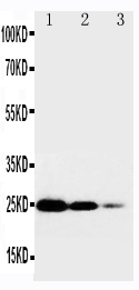
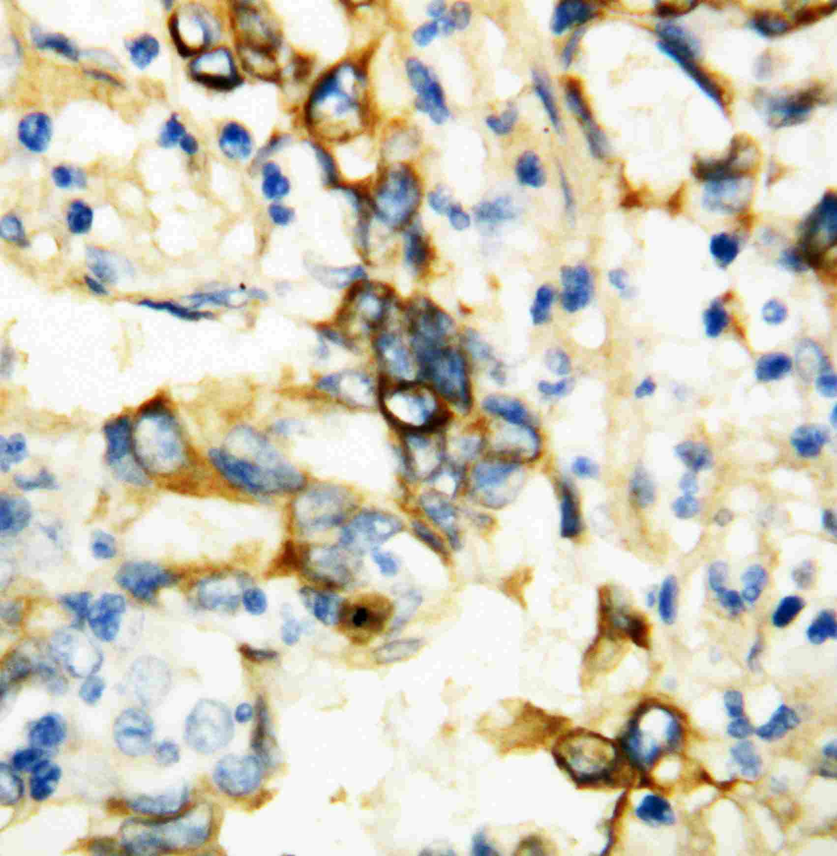
Application
| WB, IHC-P |
|---|---|
| Primary Accession | P19438 |
| Host | Rabbit |
| Reactivity | Human |
| Clonality | Polyclonal |
| Format | Lyophilized |
| Description | Rabbit IgG polyclonal antibody for Tumor necrosis factor receptor superfamily member 1A(TNFRSF1A) detection. Tested with WB, IHC-P in Human. |
| Reconstitution | Add 0.2ml of distilled water will yield a concentration of 500ug/ml. |
| Gene ID | 7132 |
|---|---|
| Other Names | Tumor necrosis factor receptor superfamily member 1A, Tumor necrosis factor receptor 1, TNF-R1, Tumor necrosis factor receptor type I, TNF-RI, TNFR-I, p55, p60, CD120a, Tumor necrosis factor receptor superfamily member 1A, membrane form, Tumor necrosis factor-binding protein 1, TBPI, TNFRSF1A, TNFAR, TNFR1 |
| Calculated MW | 50495 MW KDa |
| Application Details | Immunohistochemistry(Paraffin-embedded Section), 0.5-1 µg/ml, Human, By Heat Western blot, 0.1-0.5 µg/ml, Human |
| Subcellular Localization | Cell membrane ; Single-pass type I membrane protein . Golgi apparatus membrane ; Single- pass type I membrane protein . Secreted . A secreted form is produced through proteolytic processing. |
| Protein Name | Tumor necrosis factor receptor superfamily member 1A |
| Contents | Each vial contains 5mg BSA, 0.9mg NaCl, 0.2mg Na2HPO4, 0.05mg Thimerosal, 0.05mg NaN3. |
| Immunogen | A synthetic peptide corresponding to a sequence in the middle region of human TNF Receptor I(195-211aa CLPQIENVKGTEDSGTT) . |
| Purification | Immunogen affinity purified. |
| Cross Reactivity | No cross reactivity with other proteins |
| Storage | At -20˚C for one year. After r˚Constitution, at 4˚C for one month. It˚Can also be aliquotted and stored frozen at -20˚C for a longer time.Avoid repeated freezing and thawing. |
| Sequence Similarities | Contains 1 death domain. |
| Name | TNFRSF1A |
|---|---|
| Synonyms | TNFAR, TNFR1 |
| Function | Receptor for TNFSF2/TNF-alpha and homotrimeric TNFSF1/lymphotoxin-alpha. The adapter molecule FADD recruits caspase-8 to the activated receptor. The resulting death-inducing signaling complex (DISC) performs caspase-8 proteolytic activation which initiates the subsequent cascade of caspases (aspartate-specific cysteine proteases) mediating apoptosis. Contributes to the induction of non-cytocidal TNF effects including anti-viral state and activation of the acid sphingomyelinase. |
| Cellular Location | Cell membrane; Single-pass type I membrane protein Golgi apparatus membrane; Single-pass type I membrane protein. Secreted. Note=A secreted form is produced through proteolytic processing |

Thousands of laboratories across the world have published research that depended on the performance of antibodies from Abcepta to advance their research. Check out links to articles that cite our products in major peer-reviewed journals, organized by research category.
info@abcepta.com, and receive a free "I Love Antibodies" mug.

Provided below are standard protocols that you may find useful for product applications.
Background
Tumor necrosis factor receptor 1(TNFR1), a potent cytokine, elicits a broad spectrum of biologic responses which are mediated by binding to a cell surface receptor. Its gene is located on 12p13.2. The coding region and the 3-prime untranslated region of TNFR1 are distributed over 10 exons. There are 2 different proteins that serve as major receptors for TNF-alpha, one associated with myeloid cells and one associated with epithelial cells. Additionally, TNFR1 associates with the MADD protein through a death domain-death domain interaction. MADD provides a physical link between TNFR1 and the induction of mitogen-activated protein(MAP) kinase(e.g., ERK2) activation and arachidonic acid release. TNFR1-induced apoptosis involves 2 sequential signaling complexes. Complex I, the initial plasma membrane-bound complex, consists of TNFR1, the adaptor TRADD, the kinase RIP1, and TRAF2 and rapidly signals activation of NF-kappa-B. In a second step, TRADD and RIP1 associate with FADD and caspase-8, forming a cytoplasmic complex, complex II.
If you have used an Abcepta product and would like to share how it has performed, please click on the "Submit Review" button and provide the requested information. Our staff will examine and post your review and contact you if needed.
If you have any additional inquiries please email technical services at tech@abcepta.com.
Ordering Information
Other Products
Shipping Information
